peptide pharmacokinetics
Subcutaneous Absorption: Why Molecular Size Decides Whether a Peptide Enters Blood or Lymph
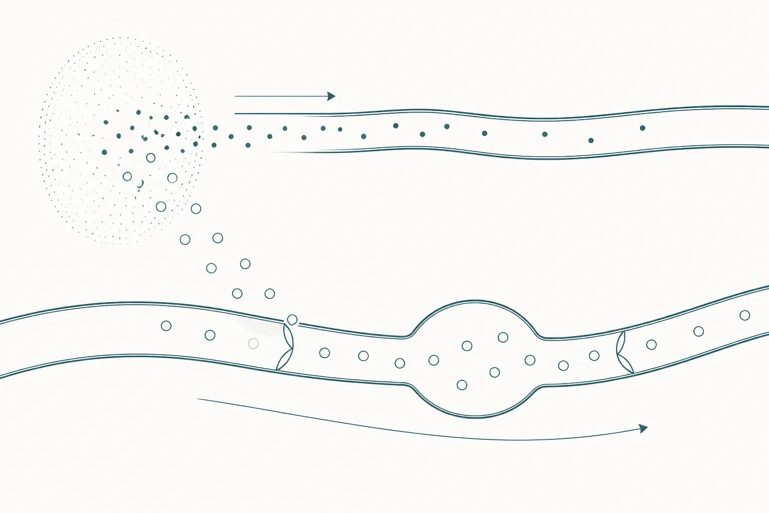
A molecule leaving a subcutaneous depot has two exits, and its size decides which one it takes. Small peptides cross the capillary wall into blood within minutes; anything much above sixteen kilodaltons travels by lymph and arrives hours later.
A molecule deposited in the subcutis has two ways out, and its size decides which one it takes. Blood capillaries are lined by a continuous endothelium whose junctions and fenestrae pass small solutes readily; the initial lymphatics are built differently, with overlapping endothelial flaps and a discontinuous basement membrane that admit macromolecules a blood capillary would exclude. The consequence is a size-dependent split of the absorbed material between two routes. In sheep, the proportion of an administered amount recovered in lymph draining the site rose roughly linearly with molecular weight across the range examined, and molecules above about sixteen kilodaltons were absorbed predominantly by the lymphatics rather than the blood 1. Most peptides of pharmacological interest sit far below that figure, which means they take the blood route almost entirely.
This article covers the entry step only. What happens to a peptide once it reaches the circulation — peptidase cleavage, glomerular filtration, and the strategies used to slow both — is the subject of the half-life and clearance article on this site. Absorption is a separate process with its own rate constant, and the distinction matters more than it sounds: for several long-acting analogues absorption is the slower of the two steps, so the observed concentration–time curve is shaped by how fast the molecule leaves the depot rather than by how fast the body destroys it 6.
Two barriers, two timescales
The subcutis is not a passive reservoir. It is a hydrated matrix of collagen and glycosaminoglycans holding interstitial fluid at a slight negative pressure, drained continuously by two independent vessel systems that differ in permeability. Blood capillaries have a continuous endothelium with tight intercellular junctions; passage across them is restricted to molecules small enough to use the paracellular pathway or the transcellular routes available to small solutes. Initial lymphatic capillaries have no continuous basement membrane, no pericytes, and overlapping endothelial cells that function as one-way flap valves opening under interstitial pressure. A macromolecule that cannot cross a blood capillary wall can still enter a lymphatic freely 2.
The two routes also operate on different timescales, and this is the part most often overlooked. Absorption into blood delivers material to the systemic circulation essentially at once. Lymph moves slowly, propelled by tissue motion and by the intrinsic contractions of collecting vessels, and it must traverse at least one lymph node before it reaches the venous system at the thoracic duct. Appearance in plasma by the lymphatic route is therefore delayed by hours rather than minutes, and the node is not merely a delay: it is a dense population of antigen-presenting cells through which the entire lymph-borne fraction is obliged to pass 4.
The molecular-weight relationship, and where peptides fall
The quantitative backbone of this field comes from cannulated-lymph experiments in sheep, in which the vessel draining the administration site is collected directly and the recovered amount measured. Four water-soluble compounds spanning two and a half orders of magnitude in molecular weight gave cumulative lymphatic recoveries that scaled with size: a few percent for a small molecule of a few hundred daltons, around a fifth for inulin at roughly five kilodaltons, approaching two-fifths for cytochrome c at about twelve kilodaltons, and close to three-fifths for a recombinant interferon of about nineteen kilodaltons. The relationship was approximately linear across that range, and the practical reading is that predominantly lymphatic absorption begins somewhere above sixteen kilodaltons 1.
| Molecule | Approximate molecular weight | Share recovered in lymph | Dominant absorption route |
|---|---|---|---|
| Small-molecule nucleoside analogue | A few hundred daltons | A few percent | Blood capillary |
| Inulin | About 5 kilodaltons | About one fifth | Blood capillary, with a lymphatic contribution |
| Cytochrome c | About 12 kilodaltons | Approaching two fifths | Mixed |
| Recombinant interferon alfa | About 19 kilodaltons | Close to three fifths | Lymphatic |
Set a peptide against that scale and the answer is immediate. A ten-residue peptide weighs a little over one kilodalton; a thirty-residue peptide around three and a half. Even a long peptide hormone is an order of magnitude below the threshold at which lymphatic uptake becomes the main route. Peptides are therefore absorbed into blood, and rapidly — which is why peptide absorption is usually discussed in terms of minutes while monoclonal antibody absorption, at around a hundred and fifty kilodaltons, is discussed in terms of days 5.
What happens between the depot and the vessel
Before a molecule reaches either vessel it has to move through the interstitium, and that transit is where a substantial fraction of the administered material is lost. The matrix is a mesh of collagen fibres and hyaluronan that hinders diffusion by size and can bind solutes by charge, so transport is partly diffusive and partly convective with bulk interstitial flow. Transit is slow enough for the tissue's own enzymes to act, and the subcutis contains peptidase activity of its own 5.
This is the standard explanation for a well-documented observation: bioavailability after subcutaneous administration is often appreciably less than complete, and the shortfall is not accounted for by anything measurable in plasma. The material is degraded before it ever arrives. For lymph-borne macromolecules there is a second opportunity for loss, because cells within the draining node can take up and catabolise a portion of what passes through. Both losses are presystemic, and neither is visible to an assay that samples only blood 24.
One implication is worth stating because it is frequently reversed. Slow absorption is not a way of protecting a molecule. A molecule that lingers in the interstitium is exposed to tissue enzymes for longer, and a construct designed to leave the depot slowly must also survive the depot. The design problem is a joint one: residence time in tissue and stability in tissue have to be solved together, not in sequence.
When absorption, not elimination, controls the curve
For a small native peptide, absorption from the subcutis is fast and elimination is fast, and the shape of the plasma curve is dominated by clearance. Acylated analogues invert this. Attaching a fatty acid gives the molecule affinity for albumin, which is the mechanism by which it escapes renal filtration, but the same chemistry also slows its departure from the depot: the acylated peptide self-associates into oligomers and binds interstitial albumin locally, and the oligomer must dissociate before absorption can proceed. Liraglutide is the documented case, where heptamer formation at the site contributes materially to the flat concentration profile, and the same principle applies to semaglutide 7.
When the absorption rate constant becomes smaller than the elimination rate constant, the terminal slope of the concentration–time curve reports absorption rather than elimination. Pharmacokineticists call this flip-flop kinetics, and it has a consequence that matters for interpreting the literature: a reported terminal half-life for such a compound is not a statement about how quickly the body destroys it. It is a statement about how quickly the depot releases it. The two can be separated only with an intravenous comparison, and where no such comparison exists the published figure remains a composite 6.
Absorption models in current use reflect this by treating the subcutaneous site as more than one compartment, with parallel first-order transfer into blood and into lymph, a dissociation step for self-associating molecules, and a presystemic loss term. Such models fit observed data well. Their parameters are largely descriptive rather than independently measured, which limits how far they can be extrapolated to a new molecule 6.
Species differences, and why the model chosen changes the answer
Almost everything quantitative in this field comes from animals, because direct lymph cannulation is not a human experiment. The sheep model that produced the molecular-weight relationship is technically excellent for exactly that reason: the vessel draining the site can be collected over many hours in a conscious animal. It is also a ruminant with a particular skin structure and lymphatic anatomy, and generalisation from it is not automatic.
A review of the methodology made the point sharply: the choice of species, the site of cannulation, the site of administration and the depth of anaesthesia each influence the quantified lymphatic transport, and reported recoveries for the same molecule differ accordingly between models 3. Anaesthesia is the most easily underestimated of these, since lymph propulsion depends in part on tissue movement, and an immobilised animal moves less lymph than a conscious one. Comparisons across published studies therefore need the model read before the number.
For acylated peptides specifically, preclinical pharmacokinetic screening has conventionally favoured minipigs over rodents, on the grounds that albumin binding and subcutaneous absorption behaviour in the pig proved more predictive of human kinetics 7. That choice is itself an admission of how strongly species affects this step: if absorption behaved uniformly across mammals, the cheapest model would do.
How to read an absorption claim
Three questions separate a well-founded statement about subcutaneous absorption from a loose one. First, what is the molecular weight, and is it anywhere near the threshold at which the lymphatic route matters? For a peptide the answer is almost always no, and any claim that a peptide is lymphatically absorbed needs the specific data. Second, was absorption measured or inferred? A lymph recovery is a measurement; an absorption rate constant fitted to a plasma curve is an inference, and a terminal half-life from a subcutaneous study alone cannot distinguish slow release from slow elimination 6. Third, in what species, under what conditions, and with what site sampled 3?
The broader point is that absorption is a genuine pharmacokinetic process rather than an administrative detail preceding the interesting part. It has its own barriers, its own losses and its own rate-limiting steps, and for the long-acting analogues that now dominate peptide pharmacology it is frequently the step that determines what the concentration profile looks like. A molecule's fate is decided as much in the tissue it starts in as in the plasma it eventually reaches.
References
- Effect of Molecular Weight on the Lymphatic Absorption of Water-Soluble Compounds Following Subcutaneous Administration
- Lymphatic transport of proteins after subcutaneous administration
- Lymphatic transport of proteins after s.c. injection: implications of animal model selection
- Subcutaneous drug delivery and the role of the lymphatics
- Mechanistic Determinants of Biotherapeutics Absorption Following SC Administration
- Pharmacokinetic Modeling of the Subcutaneous Absorption of Therapeutic Proteins
- The Discovery and Development of Liraglutide and Semaglutide