peptide pharmacokinetics
Plasma Protein Binding: Why Only the Free Fraction Acts
A peptide bound to albumin cannot engage a receptor or cross a filter. That single fact explains half-life extension by acylation — and also explains why the most common inference drawn from a protein-binding number is the wrong one.
A peptide in plasma exists in two states: associated with a plasma protein, or not. Only the unassociated portion can diffuse out of the vasculature, reach a receptor, be filtered by the kidney or be attacked by many peptidases. This is the free-drug hypothesis, and it is the single most useful idea in this part of pharmacokinetics — the concentration that matters is the unbound concentration at the target, not the total concentration in a tube 1. For acylated peptides the point is unusually concrete, because binding is not an incidental property of such molecules but the mechanism they were designed around.
The same idea is also the source of a durable error. Because the free fraction is what acts, it is tempting to conclude that a highly bound compound is a poorly performing one and that reducing binding would increase activity. That inference is wrong, and has been argued against at length in the pharmacokinetic literature 12. Understanding why requires separating two quantities that the phrase protein binding runs together: the fraction unbound, which is a ratio, and the unbound concentration, which is an amount.
The free-drug hypothesis, stated precisely
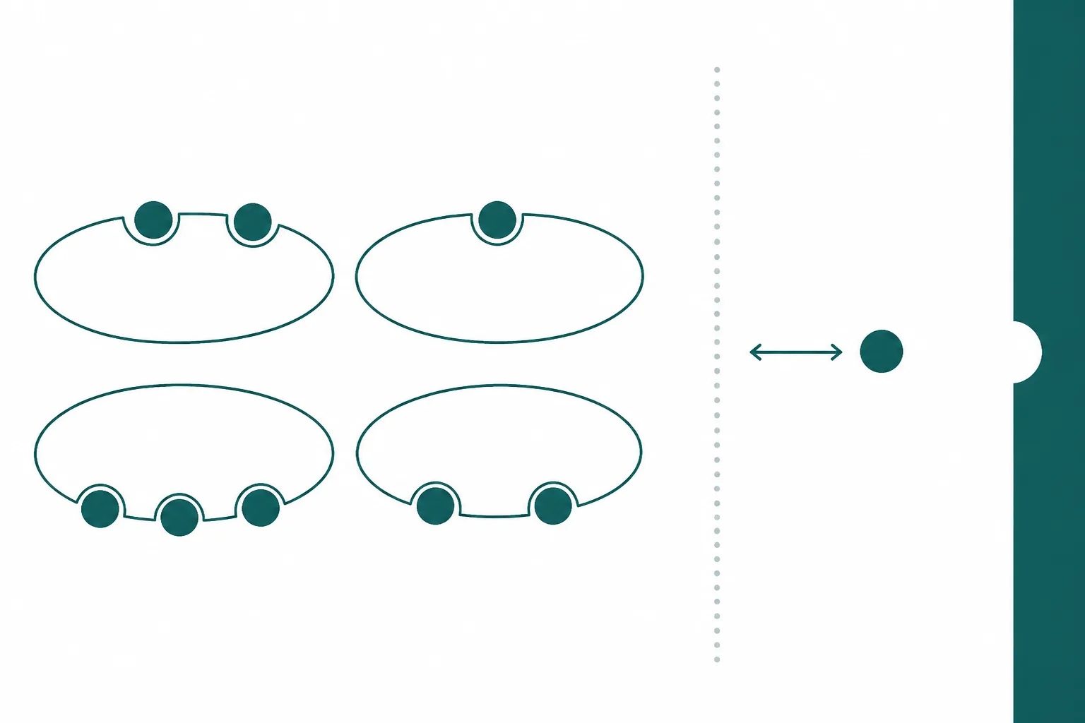
At steady state, and in the absence of active transport, the unbound concentration of a compound equilibrates across membranes. A receptor on the outside of a cell therefore sees the unbound concentration in the interstitial fluid, which tracks the unbound concentration in plasma. Bound drug is not lost, but it is not available either: it is held in a compartment from which it must dissociate before it can do anything. The hypothesis is an equilibrium statement, and it holds well for passively distributed compounds 13.
The arithmetic that follows is where intuition fails. For a compound cleared by a low-extraction process, the clearance of total drug is proportional to the fraction unbound: make the molecule less bound and the body clears it faster, in the same proportion. Total concentration falls, unbound concentration is unchanged, and the pharmacology is identical. Reducing binding has therefore bought nothing except a different-looking total-drug number. This is the core of the misconceptions argument, and it is the reason optimising a compound series towards lower protein binding is a mistake rather than an improvement 1.
The same logic disposes of the classical displacement worry — the idea that one compound displacing another from albumin produces a dangerous rise in free concentration. Displacement raises the unbound fraction transiently, clearance of unbound drug rises with it, and the system returns to the same unbound concentration at a lower total. The clinical relevance of binding changes is, on this analysis, small in most cases 2.
Why binding is the point for acylated peptides
For most small molecules, plasma protein binding is a consequence of physicochemistry that the chemist works around. For acylated peptides it is the engineering objective. A small peptide is filtered by the glomerulus essentially unrestricted, whereas albumin is retained almost completely 6. Attaching a fatty acid gives the peptide affinity for albumin's long-chain fatty acid sites, so at any instant most of the compound is attached to a carrier that the kidney will not pass. Only the unbound portion is filtered, and a binding level above ninety-nine percent reduces the filtered flux by roughly the same factor 5.
The chemistry behind that arrangement was worked out on acylated insulins, where fatty acids of different chain lengths were attached to a lysine side chain and the resulting albumin association constants measured directly. Two findings from that work shaped everything afterwards: the acylated molecules bind at albumin's long-chain fatty acid sites, and binding affinity correlated with the timing of the effect in vivo — tighter binding, more protracted action 4. Affinity is a dial connected to duration.
It is a dial with a ceiling, however, and the reason is the free-drug hypothesis running in the other direction. If affinity is too high, the free fraction becomes so small that too little peptide is available to the receptor at any moment, and potency in vivo suffers even though exposure looks excellent. Acylation design therefore aims at an intermediate affinity rather than the highest achievable one, which is part of why diacid chains — whose terminal charge tempers hydrophobic binding — displaced plain alkyl chains in later analogues 5.
Depot or sink: the difference is kinetic
Albumin binding is often described as sequestration, a word that implies loss. Whether that is accurate depends entirely on how fast the association reverses. If dissociation is rapid relative to the processes consuming free peptide, albumin behaves as a buffer: free peptide removed by a receptor or a filter is replaced immediately from the bound pool, and the plasma concentration declines slowly instead of collapsing. That is a depot, and it is the intended behaviour. If dissociation is slow, or if the peptide is covalently attached to its carrier, the bound pool is a sink and the molecule is unavailable in proportion.
The distinction is not visible in a binding percentage. Two compounds can both be ninety-nine percent bound and behave completely differently, because the number reports an equilibrium position and says nothing about the rate at which that equilibrium is maintained. Any characterisation of an albumin-binding peptide therefore needs association and dissociation kinetics alongside the affinity constant, and reviews of protein binding practice make the same point for drug candidates generally 3.
| Method | What it measures | Principal limitation |
|---|---|---|
| Equilibrium dialysis | Fraction unbound in plasma at equilibrium | Small analytical errors dominate when binding exceeds about ninety-nine percent; non-specific adsorption to the device |
| Ultrafiltration | Unbound concentration in the filtrate | Fast, but prone to adsorption and to concentration shifts during filtration |
| Surface plasmon resonance against immobilised albumin | Affinity and association and dissociation rates | Immobilised albumin is not plasma; competition from endogenous ligands is absent |
| Total-drug assay in plasma | Bound plus unbound together | For a highly bound peptide this reports mostly the depot, which is mostly inactive |
The measurement problem at high binding deserves emphasis because it is where most published uncertainty in this area lives. Distinguishing ninety-nine point five percent bound from ninety-nine point nine percent bound means resolving a free fraction that differs fivefold while the total signal barely moves. Method choice, device materials, incubation time and the plasma source all shift the answer, and cross-study comparisons of free fraction for strongly bound compounds should be treated as approximate 3.
The free fraction is not a constant
A binding figure is measured in a particular plasma, and plasma varies. Albumin concentration falls in inflammation, in liver disease, in nephrotic syndrome and in malnutrition, and rises with dehydration. Endogenous long-chain fatty acids compete for the same sites an acylated peptide uses, and their concentration changes with feeding state and with metabolic disease. Other albumin-bound compounds compete as well. The carrier, in other words, is a physiological variable rather than a fixed laboratory reagent 34.
For an acylated peptide this has a specific consequence: part of its pharmacokinetic behaviour is a property of the recipient's plasma, not of the molecule. It also means that species comparisons require care, since albumin sequence and fatty-acid binding differ between species enough that binding measured in one animal's plasma is not transferable to another's 5. Where binding is the mechanism of half-life extension, the species chosen for pharmacokinetic screening is part of the result.
Comparing in vitro with in vivo without error
The practical use of all this is extrapolation. A receptor assay reports a concentration producing a given effect in the medium of that assay; a plasma measurement reports a concentration in plasma. The two are commensurable only when both are expressed as unbound concentrations, because assay media differ enormously in protein content — a serum-free buffer binds almost nothing, while a medium containing serum binds an acylated peptide substantially. Comparing a buffer-based potency figure against a total plasma concentration is a category error, and it is common 13.
The same correction explains a frequent observation about acylated analogues: they are less potent than the native peptide in receptor assays, yet more effective in vivo. Part of the apparent potency loss is the fatty acid interfering with receptor engagement, and part is albumin in the assay medium removing the compound from solution. Disentangling the two requires measuring the unbound concentration in the assay itself, which is rarely reported 5.
The summary position is that plasma protein binding is neither good nor bad. It is a property that determines how much of a compound is available at any instant, and for peptides it is often the deliberate mechanism by which a molecule survives the kidney at all. What makes it treacherous is the ease with which a ratio gets mistaken for an amount, and a depot for a loss.
References
- The effect of plasma protein binding on in vivo efficacy: misconceptions in drug discovery
- Changes in plasma protein binding have little clinical relevance
- Plasma protein binding: from discovery to development
- Albumin binding of insulins acylated with fatty acids: characterization of the ligand-protein interaction and correlation between binding affinity and timing of the insulin effect in vivo
- The Discovery and Development of Liraglutide and Semaglutide
- Properties of the glomerular barrier and mechanisms of proteinuria