How Peripheral Peptides Reach the Brain
Most peptides do not cross the blood-brain barrier, yet some given outside the nervous system change activity deep in it. Three competing explanations have each been tested in rodents, and they do not agree.
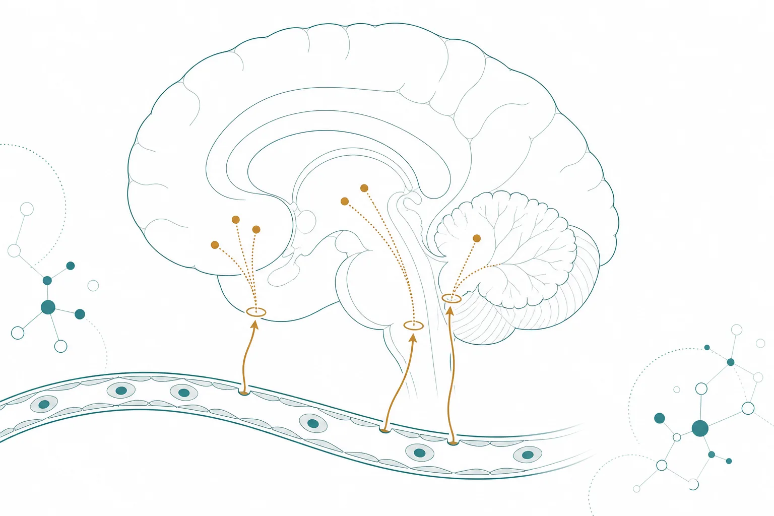
The best-supported answer is that peripherally administered peptides mostly do not cross the barrier at all; they reach neurons at the few places where the barrier is absent or specialised, and from there act on circuits that reach the rest of the brain. A second route, relay through the vagus nerve, is real but contributes less than once assumed. In rodents, mapping studies place a labelled long-acting peptide at the circumventricular organs and at selected hypothalamic sites, not throughout the parenchyma 14. Evidence tier for this article is stated per section, and almost all of it is rodent.
The question matters because the three explanations have different consequences for how a peptide's central effects should be interpreted. If access is restricted to a few sites, then the pattern of downstream activity reflects the anatomy of those sites. If vagal relay dominates, the central effect is a secondary consequence of signalling at the gut. The evidence reviewed here supports neither extreme.
The barrier and why peptides are excluded
Evidence tier for this section: anatomical and physiological review. The blood-brain barrier generally consists of endothelial tight junction barriers that prevent the free entry of blood-derived substances, which maintains the extracellular environment of the brain 7. A peptide is large and polar compared with the small lipophilic molecules that diffuse across endothelial membranes, and the junctions leave no gap between cells for it to pass. Its exclusion is the default.
That default is what makes central effects of a subcutaneously administered peptide puzzling. If the compound stays in the vasculature, something has to carry the information to neurons. The candidate explanations are access at barrier-free sites, active transport across or around the barrier, and relay through peripheral nerves.
Circumventricular organs: the gaps in the barrier
Evidence tier for this section: anatomical review of rodent and human tissue. The circumventricular organs sit along the midlines of the brain ventricles and lack the endothelial barrier, with fenestrated capillaries that transduce information between the circulation and the brain 7. The sensory ones include the organum vasculosum of the lamina terminalis, the subfornical organ and the area postrema. They have size-selective and heterogeneous vascular permeabilities, so "unprotected" overstates it: access varies by site and by molecular size.
The same review reports that neural stem cell-like cells in these organs express tight junction proteins and surround mature neurons, performing barrier functions while keeping the capacity to produce new cells 7. The gaps are therefore not holes in a wall. They are specialised interfaces with their own local protection.
Mapping studies: where a labelled peptide is found
Evidence tier for this section: rodent, mouse and rat, with labelled peptide. In rats, labelled semaglutide directly accessed the brainstem, the septal nucleus and the hypothalamus, did not cross the barrier, and interacted with the brain through the circumventricular organs and several select sites adjacent to the ventricles 1. In mice, a fluorescently labelled liraglutide injected peripherally was present in the circumventricular organs and bound neurons in the arcuate nucleus and other discrete hypothalamic sites 4.
The mouse study added a control that carries most of the weight: labelled liraglutide binding was not seen in mice lacking the receptor, so uptake in the brain depended on the receptor and was not simple tissue deposition 4. Binding at a site is evidence of access. It is not, by itself, evidence that the access causes the measured behavioural change.
| Study | Species | Finding on access | Limit |
|---|---|---|---|
| Gabery et al., 2020 | Rat | Semaglutide reached brainstem, septal nucleus and hypothalamus via circumventricular organs; did not cross the barrier | Distribution and activity maps are correlational |
| Secher et al., 2014 | Mouse and rat | Labelled liraglutide at circumventricular organs and arcuate nucleus; uptake absent without the receptor | Weight reduction in rats was independent of the receptor in vagus, area postrema and paraventricular nucleus |
| Imbernon et al., 2022 | Mouse | Tanycytes shuttled liraglutide into the hypothalamus, bypassing the barrier | Mediobasal hypothalamus only; not tested elsewhere |
Direct access against downstream activation
Evidence tier for this section: rat, activity mapping. The c-Fos protein is an activity marker, expressed in neurons that have recently been strongly stimulated. Semaglutide induced it in 10 brain areas, including hindbrain areas the drug reached directly and secondary areas with no direct receptor interaction, such as the lateral parabrachial nucleus 1. Activity in a region therefore does not mean the peptide reached it.
This matters for interpretation of any single-site result. A region that lights up could be a primary target or a synaptic downstream station. The authors combined distribution, activity, receptor location and connectivity to argue that the activation involves neurons in the lateral parabrachial nucleus controlling meal termination 1. That is an inference from convergence of maps, and not a demonstration of causation.
The vagal relay model, and what tests of it found
Evidence tier for this section: rat and mouse, surgical and genetic manipulation. In rats with complete subdiaphragmatic vagal deafferentation, higher doses of two long-acting agonists were needed to suppress food intake, which was observed at later time points only. Blocking central receptors by ventricular delivery of an antagonist also attenuated the effect in intact rats. The authors concluded that both vagal afferent receptors and direct central receptor activation mediate the intake suppression 5.
A mouse study approached the question from the central end. Preproglucagon neurons in the nucleus of the solitary tract are widely assumed to link the gut and brain GLP-1 systems. They were found to receive vagal input mainly from oxytocin-receptor-expressing neurons, not from those expressing the GLP-1 receptor, and were not necessary for eating suppression by agonist drugs. The authors concluded that central and peripheral GLP-1 systems act through independent circuits 2.
Ablation experiments and their contradictions
Evidence tier for this section: rodent, with conflicting results. In rats, liraglutide-dependent body weight reduction was independent of the receptor in the vagus nerve, the area postrema and the paraventricular nucleus 4. The vagal deafferentation study found that the vagus mattered for potency 5. These are not necessarily contradictory, because one removed the receptor at a site and the other removed the whole afferent pathway, and the outcomes differed. They are not reconciled either.
A review of the brain GLP-1 literature notes that evidence for area postrema involvement is mixed: one study found that lesions reduced feeding suppression after portal GLP-1 infusion, another found no effect on liraglutide 3. The same review concludes that if these drugs act partly through the central nervous system, they likely reach the receptors directly, because the relevant neurons do not respond to the endogenous gut hormone in the way a relay would predict 3. This is a reviewer's inference.
Transport routes: tanycytes and the minority pathways
Evidence tier for this section: mouse. Tanycytes are specialised ependymoglial cells lining the third ventricle. Liraglutide was shuttled to target cells in the mouse hypothalamus by tanycytes, bypassing the barrier. Selectively silencing the receptor in tanycytes, or inhibiting tanycytic transcytosis with botulinum neurotoxin expression, hampered the drug's transport into the brain and its activation of hypothalamic neurons, and blocked its reported effects on food intake, body weight and fat mass 6.
This is the clearest example in the cited literature of a receptor-dependent route that is neither passive diffusion nor vagal relay. Other carrier-mediated and receptor-mediated transport systems are described in textbook physiology for particular peptides, but none is examined in the sources reviewed here, and this article does not extend the tanycyte result to peptides generally.
Why rodent data do not transfer cleanly to human brains
Evidence tier for this section: methodological. Every mapping, ablation and transport study cited above was done in mice or rats, with labelled compound, tissue sectioning or genetic manipulation that cannot be performed in people 146. None reports the distribution of a labelled peptide in the human brain. The anatomy of the circumventricular organs is conserved in outline, but the relative size of each organ, the density of fenestrated vessels and the extent of tanycytic contact differ between species.
The step from rodent to human is also a step from labelled tissue to indirect measures. Human evidence on central access of peripheral peptides comes mainly from imaging of activity and from behavioural and physiological endpoints, which show that something changed centrally without showing where the molecule went.
What this means for a researcher reading the literature
Three checks apply to any claim that a peptide "crosses the blood-brain barrier" or "acts centrally". Ask whether the study showed the molecule at a site, or only showed activity at a site. Ask whether a barrier-free organ, a transport cell or a nerve relay was tested as the route. And ask whether the species and compound match the one the claim is about. Binding at a circumventricular organ, activation downstream, and a behavioural change are three separate observations.
The current picture is of several partly independent routes, each demonstrated in rodents with a specific long-acting agonist. Whether the same routes operate for a different peptide, in a different species, at a different exposure, is a question the existing mapping studies were not designed to answer.
References
- Semaglutide lowers body weight in rodents via distributed neural pathways
- Central and peripheral GLP-1 systems independently suppress eating
- The diverse effects of brain glucagon-like peptide 1 receptors on ingestive behaviour
- The arcuate nucleus mediates GLP-1 receptor agonist liraglutide-dependent weight loss
- Peripheral and central GLP-1 receptor populations mediate the anorectic effects of peripherally administered GLP-1 receptor agonists, liraglutide and exendin-4
- Tanycytes control hypothalamic liraglutide uptake and its anti-obesity actions
- New aspects in fenestrated capillary and tissue dynamics in the sensory circumventricular organs of adult brains