receptor pharmacology fundamentals
Biased Agonism: G-Protein Versus β-Arrestin Signalling at Peptide Receptors
Two agonists can occupy the same receptor and produce different mixtures of downstream signal. That observation dismantled the idea of a receptor as an on-off switch, and it is the reason two compounds with identical targets can behave differently over time.
Biased agonism is the observation that two agonists binding the same receptor can produce different proportions of the downstream signals that receptor is capable of generating. One ligand may drive G-protein coupling strongly and recruit β-arrestin weakly; another may do the reverse; a third may activate both in the ratio the endogenous hormone does. The effect at the receptor is not a matter of how much signal, but of which mixture of signals, and that distinction is invisible to any assay that measures a single readout 12.
The concept matters for peptide pharmacology specifically. Most of the receptors that peptide hormones act on are G-protein-coupled receptors, the same family in which bias was first characterised, and several of the best-documented examples come from the incretin and melanocortin systems. It also supplies the mechanism behind an otherwise confusing fact: two compounds described as agonists at the same target, with comparable potency, can differ in how long their effect persists and in which adverse effects accompany it.
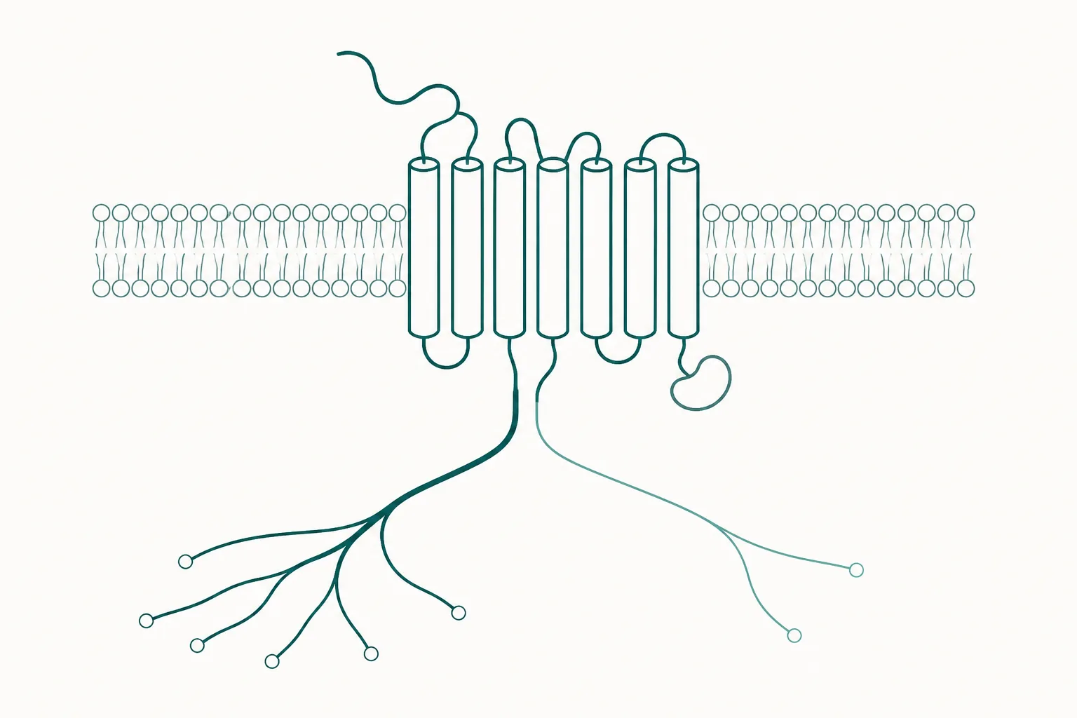
Why a receptor is not a switch
The classical account of receptor activation has two states. The receptor is either unoccupied and silent, or occupied and active, and an agonist's job is to shift the population towards the active state. On that account the only meaningful differences between two agonists are how tightly each binds and how much response each can produce at saturation. Both of those are single numbers, and both were measured for decades before anything else was suspected.
What replaced it is a model in which the receptor adopts many conformations, each coupling with different efficiency to different intracellular partners, and in which a ligand stabilises a particular subset of those conformations rather than flipping a switch. The receptor becomes, in the phrase used in one influential review, less a switch than an allosteric processor that converts the structural information in a bound ligand into a pattern of outputs 5. Two ligands stabilising different conformational subsets therefore deliver different patterns, and the size of that difference is what the word bias names.
The two arms, and what each one does
The G-protein arm is the one described in every textbook account. Agonist binding promotes coupling to a heterotrimeric G protein, which dissociates and acts on effector enzymes — adenylyl cyclase for Gs, phospholipase C for Gq — producing second messengers within seconds. This arm generates the response most assays are built to detect, because cyclic AMP and intracellular calcium are convenient to measure.
The β-arrestin arm was discovered as a braking mechanism and turned out to be more than that. After activation, G-protein-coupled receptor kinases phosphorylate the receptor's intracellular regions, and β-arrestin binds the phosphorylated receptor, sterically blocking further G-protein coupling and recruiting the machinery that draws the receptor into the cell. That much is desensitisation. The additional finding was that β-arrestin also functions as a genuine adaptor protein, assembling cytoplasmic signalling complexes and transducing signals to effector pathways in its own right, with biochemical and functional consequences distinct from those of the G-protein arm 3.
Because the same protein performs both the switching-off and a signalling function, arrestin recruitment has two separable consequences for any agonist that produces it. It shortens the G-protein signal and it opens a second output. A compound that recruits arrestin poorly will tend to sustain the G-protein signal for longer and to generate less of the arrestin-dependent output, and those two effects arrive together whether or not both are wanted 2.
| G-protein arm | β-arrestin arm | |
|---|---|---|
| Immediate event | Coupling to a heterotrimeric G protein | Receptor phosphorylation, then arrestin binding |
| Typical timescale | Seconds | Seconds to minutes, after the G-protein signal |
| Common readout | Cyclic AMP, intracellular calcium | Arrestin recruitment assays, receptor internalisation |
| Effect on the other arm | Triggers the kinases that recruit arrestin | Blocks further G-protein coupling |
| Second role | Effector enzyme activation | Scaffolding of independent signalling complexes |
| Consequence for receptor location | Receptor remains at the surface | Receptor is internalised, then recycled or degraded |
What bias means, and what it does not
Three things get called bias in the literature and only one of them is a property of the ligand. Ligand bias is the genuine article: a difference in the relative pathway activation produced by two ligands at the same receptor in the same system. System bias is a property of the cell, which may express one transducer abundantly and another sparsely, so that the pathway with more amplification appears dominant regardless of the ligand. Observational bias is a property of the assay, which may sample one pathway at a timepoint that flatters it 1.
Only relative measurements survive those confounds. A claim that a compound is a G-protein-biased agonist means nothing without a named comparator, because bias has no absolute scale. The comparator is normally the endogenous ligand, and the claim is then that the test compound departs from the natural signalling ratio in a stated direction 1.
How bias is quantified
The standard approach rests on an operational model that describes agonism using two parameters: the affinity of the ligand for the receptor, and its efficacy in activating a particular transducer. Combining them gives a transduction coefficient for each pathway, usually expressed as the logarithm of the efficacy-to-affinity ratio. Taking the difference between a ligand's coefficients for two pathways, and then subtracting the same difference calculated for a reference agonist, yields a bias factor in which the contribution of the cell system cancels out 4.
- Measure full concentration–response curves for the test ligand in each pathway of interest. Partial curves cannot support the calculation.
- Fit the operational model to obtain a transduction coefficient per pathway.
- Repeat for a reference agonist, normally the endogenous ligand, in the same cell background.
- Take the difference of differences. What remains is attributable to the ligand rather than to the assay.
- Report the comparator, the pathways, the cell background and the timepoints. Without them the number is uninterpretable.
Two limitations deserve stating plainly. The first is kinetic: pathway measurements are taken at different times after agonist addition, second messengers accumulating within seconds while arrestin recruitment and internalisation unfold over minutes, and a ligand that dissociates quickly can appear biased purely because of when the readings were taken 5. The second is that most measurements are made in cell lines overexpressing the receptor, where the stoichiometry between receptor and transducer bears no necessary relation to that of any native tissue.
The GLP-1 receptor as a worked example
The incretin field provides the most developed peptide example. Work on agonists at the GLP-1 receptor reported that compounds which retained the receptor at the plasma membrane produced greater long-term insulin release from beta cells, and that this outcome depended on reduced β-arrestin recruitment together with faster dissociation of the agonist from the receptor. In mouse models the same compounds improved glycaemic measures without the accompanying behavioural signs that accompany conventional agonists at this receptor 6.
The mechanism proposed there is instructive because it is not simply more signal. Less arrestin recruitment means less internalisation, which means more receptor available at the surface over an extended period, which changes the shape of the response over hours rather than its peak height. A single-timepoint potency measurement would rank these compounds differently from a sustained-release measurement, and the two rankings are both correct about different things.
A second example from the same receptor family concerns the dual agonist tirzepatide, which engages both the GIP receptor and the GLP-1 receptor. Its characterisation reported that engagement of the two receptors is unequal, and separately that its signalling at the GLP-1 receptor is biased, with weaker β-arrestin recruitment relative to what native GLP-1 produces at that receptor 7. Whether that bias contributes to the compound's clinical profile has not been established. It is a mechanistic hypothesis with in vitro support, and it is routinely reported as more than that.
The melanocortin example
The melanocortin-4 receptor supplies a second case, and a structurally resolved one. Cryo-electron microscopy structures of the active receptor bound to two peptide agonists and coupled to Gs showed how each ligand sits in the binding pocket and which helix movements follow, and identified one of the two — a cyclic peptide agonist used clinically in rare genetic disorders of the melanocortin pathway — as biased towards Gq/11 signalling 8. The structural work therefore attaches a signalling preference to a specific set of ligand-receptor contacts rather than leaving bias as a purely functional observation.
This matters for the family generally because melanocortin receptors are distributed across tissues with different physiological jobs, and because earlier agonists at this receptor were associated with cardiovascular effects that later compounds were not. A signalling explanation for such a difference is attractive and partially supported. It is worth noting that a selectivity explanation and a bias explanation are both available for the same observation, and distinguishing them requires data that does not exist for most compounds in this class.
Why bias is invoked for tolerance and for side effects
The therapeutic argument for biased ligands has one clear shape. If the wanted effect of a receptor runs through one arm and an unwanted effect runs through the other, a ligand that separates them is better than one that does not, regardless of potency. Since arrestin recruitment also drives the internalisation that reduces responsiveness over time, an arrestin-sparing agonist may in principle sustain its effect longer than one that recruits arrestin efficiently 25.
That argument has held up unevenly. Its clearest failure mode is the assumption that each effect belongs to one arm, which is often not the case: both arms feed into overlapping networks, and the same pathway can serve wanted and unwanted outcomes in different tissues. The general finding after two decades of work is that bias is real, measurable and mechanistically informative, and that the step from a bias factor in a cell line to a predicted clinical advantage in a person remains the weakest link in the chain 15.
What the evidence does not establish
- That a bias factor measured in a transfected cell line predicts the behaviour of the same compound in native tissue, where receptor and transducer abundance differ.
- That an arrestin-sparing profile confers durable clinical benefit. The proposition is reasonable and has preclinical support; controlled human comparisons designed to test it are scarce.
- That the two-arm description is complete. G-protein subtypes, receptor dimerisation, compartmentalised signalling from endosomes and receptor-associated proteins all contribute outputs that a two-pathway measurement cannot see.
- That a compound marketed as biased has been characterised as such. Bias requires a named comparator, full curves in both pathways and a stated cell background; the label is frequently applied without any of them.
Why this framework is worth holding
Biased agonism is the concept that makes several otherwise puzzling observations in peptide pharmacology legible at once. It explains how two agonists at one receptor can differ in the persistence of their effect rather than only in its size. It explains why a potency figure measured on one readout does not rank compounds reliably on another. It explains why the receptor's location inside the cell, rather than merely its occupancy, is part of the pharmacology 6.
What it does not supply is a shortcut. Establishing that a compound is biased requires more experimental work than establishing that it is an agonist, and translating a bias profile into a prediction about a living system requires more still. The defensible position is that the framework is well founded, that its measurements are meaningful when fully reported, and that the inferential distance between a signalling ratio in a dish and an outcome in an organism remains large 1.
References
- Signalling bias in new drug discovery: detection, quantification and therapeutic impact
- Teaching old receptors new tricks: biasing seven-transmembrane receptors
- Transduction of receptor signals by beta-arrestins
- A simple method for quantifying functional selectivity and agonist bias
- Biased signalling: from simple switches to allosteric microprocessors
- Targeting GLP-1 receptor trafficking to improve agonist efficacy
- Tirzepatide is an imbalanced and biased dual GIP and GLP-1 receptor agonist
- Structures of active melanocortin-4 receptor-Gs-protein complexes with NDP-α-MSH and setmelanotide