Kisspeptin-10 and KISS1R: Potency and Half-Life
The ten-residue C-terminal fragment binds the receptor about as well as the fifty-four-residue parent. In rodents it does not behave that way, and the resolution turns out to be only partly pharmacokinetic.
Kisspeptin-10 is an agonist at KISS1R, a Gq/11-coupled G protein-coupled receptor, and it is not a separate molecule from kisspeptin-54 so much as the active end of it: both are products of the KISS1 gene, and the shorter peptide is the parent's C-terminal decapeptide. When the kisspeptins were first isolated from human placenta and matched to what was then the orphan receptor GPR54, the 54-, 14- and 13-residue forms all bound rat and human receptor with low nanomolar affinity and all triggered the same downstream events — phosphoinositide hydrolysis, calcium mobilisation, ERK1/2 and p38 phosphorylation 1. That is an in vitro result, and it is where the agreement between the two peptides ends.
In rodents given the peptides systemically, the 54-residue form sustains luteinising hormone release far longer than the decapeptide 6. In the one published human study that infused both against a common comparator, they produced similar gonadotrophin responses 5. The two literatures therefore disagree, and the disagreement is informative rather than embarrassing: working out why has produced a cleaner account of what limits a short peptide in a whole animal than either result would have given alone.
The receptor sits upstream of GnRH, not at the pituitary
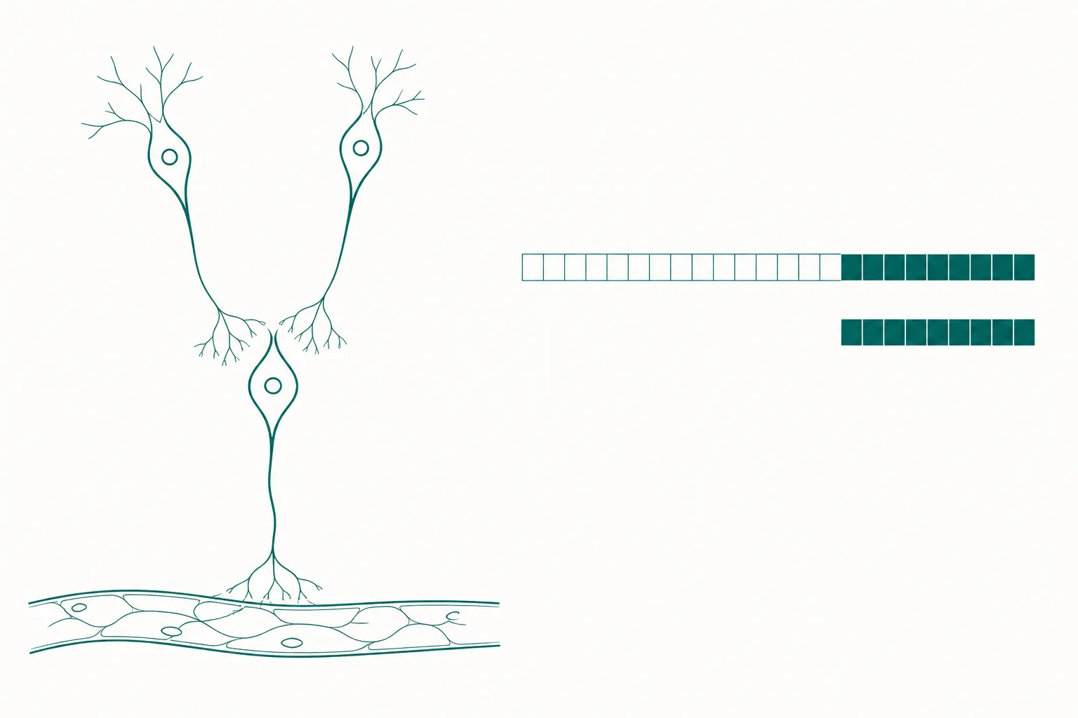
Where a receptor sits in a cascade determines what activating it can and cannot do, and for KISS1R that position was settled by genetics rather than by pharmacology. Two groups reported in 2003 that loss of function in the gene then called GPR54 causes isolated hypogonadotropic hypogonadism — a failure of pubertal development with intact pituitary and hypothalamic anatomy. One studied a consanguineous family in which affected members were homozygous for an L148S substitution, with a second unrelated proband carrying two further mutations; transfected cells expressing the mutant receptors accumulated significantly less inositol phosphate than wild type 2. The other mapped a new locus to chromosome 19p13 in a large consanguineous family with five affected siblings, all homozygous for a 155-nucleotide deletion spanning an intron-exon junction 3.
The mouse arm of the first study is what localises the lesion. Receptor-deficient mice had isolated hypogonadotropic hypogonadism, but they responded normally to both exogenous gonadotropins and exogenous GnRH, and their hypothalamic GnRH content was normal 2. The machinery below the receptor was therefore intact and stocked; what had failed was the signal to release. This is human and mouse genetic evidence, not a pharmacological demonstration, and it carries a specific implication for any agonist at this receptor: the response depends on an intact GnRH neuron population downstream, so the ceiling is set by that population rather than by receptor occupancy.
Why the decapeptide retains binding
The kisspeptins isolated from placenta came in 54-, 14- and 13-residue lengths sharing a common RF-amide C terminus, and all bound the receptor at low nanomolar concentrations 1. The shared C-terminal region is the pharmacophore; the additional residues of the longer forms are not required for receptor engagement. This is the same active-fragment pattern that truncation experiments established for growth hormone releasing hormone, and it recurs often enough in peptide endocrinology to be worth stating as a general expectation: a peptide hormone frequently carries its receptor-binding information in a minority of its chain.
What the pattern does not license is the inference that the fragment will therefore substitute for the parent in an animal. Binding affinity is measured in a dish, at a defined concentration, against a receptor that the experimenter has already placed in front of the ligand. An intact organism adds proteolysis, distribution, clearance and anatomical barriers, none of which the assay interrogates. The kisspeptins are a clean demonstration that those additional terms can dominate.
The divergence in vivo, measured in mice
A 2017 study administered each peptide systemically to male mice and sampled plasma luteinising hormone at ten minutes and at two hours. The longer form sustained hormone release far beyond the point at which the decapeptide's effect had subsided, which the authors took as evidence of a differential mode of action rather than a difference in degree. Measured in the same animals, plasma half-life was approximately 32 minutes for the 54-residue peptide and approximately 4 minutes for the decapeptide 6. These are mouse figures and apply to mice.
An eightfold difference in half-life is an obvious candidate explanation, and the study then tested it directly rather than assuming it. Repeated injections of the decapeptide every ten minutes over an hour — an attempt to hold exposure up by brute force — failed to reproduce the sustained hormone rise produced by a single injection of the longer peptide 6. That is a negative result against the simplest hypothesis, and it is the most useful experiment in the paper.
The human comparison, and its sample size
Human work on these peptides began with a double-blind, placebo-controlled crossover study in six male volunteers, each receiving a 90-minute intravenous infusion of the 54-residue peptide and a saline control in random order. Luteinising hormone, follicle-stimulating hormone and testosterone all rose significantly against saline; mean luteinising hormone over the 90 minutes was 10.8 ± 1.5 U/l on peptide against 4.2 ± 0.5 U/l on saline 4. That established that the axis responds in humans. It did not compare the two isoforms.
The comparison came in 2015, in a single-blinded placebo-controlled physiological study in healthy men. Participants received vehicle, kisspeptin-10, kisspeptin-54 and GnRH intravenously over three hours on separate study days at least a week apart, each at three ascending matched infusion rates, with five participants per group. At the highest rate, mean area under the curve for serum luteinising hormone during infusion was 10.81 ± 1.73 h.IU/l for the decapeptide, 14.43 ± 1.27 h.IU/l for the 54-residue peptide and 34.06 ± 5.18 h.IU/l for GnRH 5. GnRH was roughly threefold higher than the decapeptide and roughly twofold higher than the longer peptide, both differences significant. Between the two kisspeptins the authors concluded that, at the rates tested, either isoform had similar effects on reproductive hormone secretion.
Three features of that study constrain how far its conclusion travels. The authors name the first themselves: the sample size was small. The second is the qualifier "at the doses tested" — a comparison at three matched rates establishes similarity over that range and not beyond it. The third is the route: continuous intravenous infusion over three hours is close to the brute-force compensation that failed in mice, and it is the administration pattern least likely to expose a difference driven by clearance. A study designed this way can return "similar" for a pair of compounds that would separate under single-bolus administration.
| Finding | Evidence tier | What it establishes |
|---|---|---|
| Both isoforms bind KISS1R at low nanomolar concentrations and activate the same pathways | In vitro, recombinant cells | The decapeptide is the pharmacophore; added residues are not needed for receptor engagement |
| Receptor loss of function causes isolated hypogonadotropic hypogonadism | Human genetics plus mouse knockout | The receptor acts upstream of GnRH release, with downstream machinery intact |
| The 54-residue form sustains luteinising hormone far longer after systemic administration | Animal model, male mice | The two peptides are not interchangeable in a whole animal |
| Plasma half-life approximately 32 minutes against approximately 4 minutes | Animal model, male mice | A clearance difference exists, in mice, of roughly eightfold |
| Repeated short-interval administration failed to reproduce the sustained response | Animal model, male mice | Clearance alone does not account for the divergence |
| Only the longer form activated c-FOS in GnRH neurons behind the barrier | Animal model, male mice | Consistent with differential blood-brain barrier access |
| Similar gonadotrophin responses to the two isoforms at three matched infusion rates | Human physiological study, five per group | No detectable difference under continuous infusion, over the range tested |
Longer-acting agonists, and a second pharmacokinetic surprise
If the limitation on the short peptide were purely one of persistence, the design response would be a receptor agonist with a longer half-life. One such compound, MVT-602, was compared against the 54-residue native peptide in healthy women in the early follicular phase, with nine participants in the main comparison, alongside cell-based signalling assays and recordings from GnRH neurons in brain slices 7. The pharmacokinetic result was not what the premise predicted: the two had closely similar half-lives, in the range of 1.68 to 2.02 hours, which the authors describe as surprisingly similar. The same paper cites a terminal half-life of approximately 4 minutes for the decapeptide from earlier human work.
The implication runs in the same direction as the failed mouse compensation experiment. Where two agonists at this receptor share a half-life and differ in the duration of the response they produce, the difference cannot be attributed to exposure, and the explanation has to be sought at the receptor — in how long and in what manner the activated receptor keeps signalling. That is a different class of question from clearance, and the literature on it for this receptor is small.
What is established, and how large the human dataset actually is
Three things are established with reasonable confidence. The receptor is required for normal gonadotrophin physiology, by convergent human and mouse genetics 23. Both principal isoforms engage it in vitro at comparable concentrations 1. Administering the longer isoform to humans raises luteinising hormone, follicle-stimulating hormone and testosterone against placebo 4. Beyond that, the evidence thins quickly, and the distinctions between tiers do real work: the sustained-release difference between the isoforms is a mouse finding, the barrier-access explanation is a mouse finding, and the human comparison that found them similar used an infusion design and five participants per group.
It is worth being explicit about the scale. Summing the human studies cited here gives six men in the first infusion study, five per group across four conditions in the comparison, and nine women in the main arm of the longer-acting agonist study. The whole human record for these peptides is measurable in tens of participants, all in controlled physiological experiments with hormone concentrations as endpoints. No clinical outcome has been established for any kisspeptin in any population, and nothing in this literature supports a numerical regimen for anything.
How to read this literature
The practical filter is to check which system produced each number before accepting any comparison between the two peptides. A statement that the longer form is more potent is a rodent statement; a statement that they are equivalent is a human statement obtained under continuous infusion; a statement that they bind comparably is an in vitro statement. All three are supported, and they are not in conflict once the system is attached to each. They look contradictory only when the system is dropped, which is how most secondary accounts of this pair are written.
The broader lesson is the one the fragment story keeps teaching. Retained binding by a truncated peptide is a statement about the receptor interaction and nothing else, and the properties that decide whether a fragment is useful in an organism — resistance to proteolysis, clearance rate, tissue access — are carried by parts of the molecule the binding assay was never asked about. Kisspeptin-10 binds its receptor essentially as well as its parent and does not substitute for it in a mouse. Both facts are correct, and keeping them in the same sentence without collapsing them is most of what reading this field well requires.
References
- The metastasis suppressor gene KiSS-1 encodes kisspeptins, the natural ligands of the orphan G protein-coupled receptor GPR54
- The GPR54 gene as a regulator of puberty
- Hypogonadotropic hypogonadism due to loss of function of the KiSS1-derived peptide receptor GPR54
- Kisspeptin-54 stimulates the hypothalamic-pituitary gonadal axis in human males
- Direct comparison of the effects of intravenous kisspeptin-10, kisspeptin-54 and GnRH on gonadotrophin secretion in healthy men
- Mechanistic insights into the more potent effect of KP-54 compared to KP-10 in vivo
- Kisspeptin receptor agonist has therapeutic potential for female reproductive disorders