structure and modification
Disulfide Bonds and Why Some Peptides Have a Shape at All
Most short peptides have no fixed structure. The ones that do usually owe it to a covalent bridge between two cysteines, and that bridge is both the reason they work and the first thing to fail.
A peptide of ten or twenty residues generally has no persistent three-dimensional structure in water. It samples conformations continuously and adopts a defined shape only on binding, if at all. The exceptions are almost all peptides containing cysteine, because cysteine can form the one covalent crosslink available within a normal amino acid repertoire. A disulfide bond converts a floppy chain into a constrained loop, and for a short hormone or toxin that single bond is frequently the difference between a molecule with a function and a molecule with none 3.
Cysteine Oxidation and How the Bond Forms
Cysteine carries a thiol side chain with a pKa near 8.3 in a free amino acid, lower in many protein environments. Deprotonation gives a thiolate anion, and it is the thiolate rather than the neutral thiol that is chemically active. Two thiolates oxidised, most simply by dissolved molecular oxygen and often catalysed by trace transition metals, form a sulfur-sulfur bond and release two hydrogens. The bond energy is around 60 kilocalories per mole, comparable to a carbon-carbon single bond, so the linkage is genuinely covalent and not a weak association.
Because the reactive species is the anion, everything about disulfide chemistry is pH-dependent in the same direction. Formation is slow in acid and fast in mild alkali. So is the reverse reaction, and so is exchange between existing bonds. A single parameter therefore governs both the synthesis of the bond and its principal degradation route, which is the central practical fact of working with cysteine-containing peptides.
The question of which cysteines pair with which is not trivial. A peptide with six cysteines admits fifteen distinct pairing arrangements; with eight, the number rises to one hundred and five. Ordinarily only one of those is biologically active. The demonstration that a fully reduced, denatured protein could reoxidise spontaneously to recover the correct pairing and full enzymatic activity established that the information specifying the fold, including its disulfide connectivity, is contained entirely in the amino acid sequence 1. That result underpins how synthetic cysteine-rich peptides are folded to this day: the chain is assembled reduced, then oxidised under conditions permissive enough for incorrect pairings to reshuffle toward the thermodynamic minimum.
Why Oxytocin and Vasopressin Need Theirs
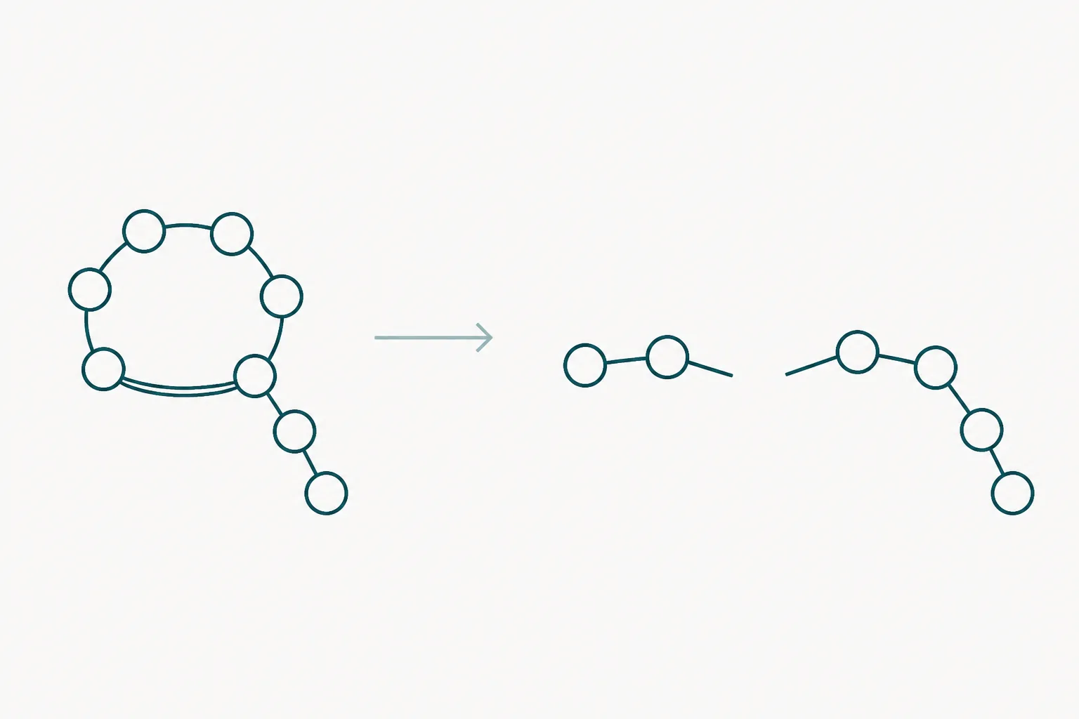
Oxytocin and vasopressin are the classical illustration because they are so small that the disulfide has nowhere to hide. Each is nine residues. In each, the cysteines at positions 1 and 6 are bridged, producing a six-residue macrocycle with a three-residue tail extending from it. The two hormones differ from one another at only two positions, yet they act at different receptors with different physiological consequences, which shows how little sequence variation a constrained scaffold needs in order to encode distinct function.
The total synthesis of oxytocin, achieved in 1953, was the first chemical synthesis of a peptide hormone and it depended on getting that ring closed correctly; the synthetic material reproduced the biological activity of the isolated hormone, which settled the structure 2. Reducing the bridge opens the ring, and the linear nonapeptide is essentially devoid of the parent activity. The residues have not changed. The topology has, and the receptor reads topology.
The same architecture recurs wherever a small peptide must hold a shape. Somatostatin uses a single disulfide to close a fourteen-residue chain into a large loop. Insulin uses three, two of them joining separate chains, which is why reduced insulin chains do not spontaneously reassemble efficiently and why the molecule is biosynthesised as a single precursor that folds first and is cut afterwards. Venom peptides push the principle furthest: cone snail and spider toxins are built on rigid cystine frameworks of two, three or four bridges, and it is that rigidity, not any unusual sequence, that gives them ion channel selectivity and remarkable stability outside the organism 5.
Scrambling: Degradation Without Fragmentation
The characteristic failure of a disulfide-containing peptide is not cleavage of the backbone. It is rearrangement of the bridges. A single thiolate anion, supplied by a free cysteine, a trace of reducing impurity, or generated in situ, attacks an existing disulfide, displaces one of its partners and creates a new free thiolate that goes on to attack the next bond. The process is catalytic: one nucleophile can reshuffle an entire population. Where more than one disulfide exists, the result is a mixture of connectivity isomers, most of them inactive 3.
What makes this the most dangerous degradation route is that it is silent to the most common analytical check. A scrambled isomer has exactly the same atoms in exactly the same numbers as the native form. Its intact mass is identical, its ultraviolet absorbance is identical, and an elemental analysis cannot separate them. Only methods sensitive to shape or to connectivity will see the difference, and reversed-phase chromatography usually will, because the isomers differ in exposed hydrophobic surface and elute at different times.
A second, slower route operates under alkaline or thermal stress. Cystine can undergo beta-elimination to give a dehydroalanine residue and a persulfide, which decomposes to release free thiol. That newly liberated thiol then catalyses scrambling, so alkaline stress both damages the bond directly and generates the catalyst for further damage. The dehydroalanine can subsequently react with a nearby amine or thiol to form an irreversible crosslink, at which point the molecule is not recoverable by any reoxidation step.
Reducing Agents, and What They Destroy
Deliberate reduction is routine in analysis and destructive to activity. Dithiothreitol reduces disulfides through a two-step thiol-disulfide exchange driven by formation of a stable six-membered cyclic disulfide, and works best above pH 7 where its own thiols are deprotonated. Tris(2-carboxyethyl)phosphine reduces by a different mechanism that does not require a thiolate, so it remains effective in acid, is odourless, and does not interfere with thiol-reactive alkylating reagents. 2-Mercaptoethanol is the older and weaker option, requiring large excess.
Biology maintains the same distinction geographically. Extracellular fluid and plasma are oxidising environments in which disulfides are stable, while the cytosol is held strongly reducing by millimolar glutathione, in which disulfides are rapidly cleaved. This is the reason disulfide-constrained peptides are robust in circulation and a poor structural choice for anything intended to act inside a cell, and it is also the mechanism exploited by disulfide-linked drug conjugates designed to release their payload after internalisation.
| Agent or condition | Effect on a disulfide | Reversible by reoxidation |
|---|---|---|
| Dithiothreitol | Complete reduction above neutral pH | Yes, if scrambling is controlled |
| Tris(2-carboxyethyl)phosphine | Complete reduction, effective in acid | Yes, if scrambling is controlled |
| Glutathione at cytosolic concentration | Reduction | Not in that environment |
| Trace copper or iron with dissolved oxygen | Catalyses oxidation and scrambling | Partially, with losses |
| Alkaline pH with heat | Beta-elimination and irreversible crosslinking | No |
| Benzyl alcohol as a preservative | None; it is not a reducing agent | Not applicable |
Mapping the Bonds, and What It Implies for Storage
Establishing which cysteines are actually paired is a defined analytical exercise. The classical approach digests the peptide enzymatically under non-reducing conditions and at mildly acidic pH, chosen specifically to suppress thiolate formation and prevent the analysis from creating the scrambling it is meant to measure. Fragments that remain joined after digestion are disulfide-linked, and their combined masses identify the pairing. Parallel analysis of a reduced and alkylated sample provides the comparison: peaks that shift are bridged, peaks that do not are free 4.
Supporting techniques include partial reduction with stepwise alkylation, which assigns bridges in order of accessibility, and quantification of free thiol using Ellman's reagent, which reports how many cysteines were never oxidised at all. Fragmentation methods based on electron transfer preferentially cleave the sulfur-sulfur bond in preference to the backbone, which allows connectivity to be read directly from an intact molecule rather than reconstructed from digest fragments, and this has become the reference approach for cysteine-rich peptides 4.
The storage implications follow from the chemistry without further argument. Every factor that raises thiolate concentration accelerates scrambling: alkaline pH, elevated temperature, dissolved oxygen, and trace transition metals, which is why chelating agents appear in many formulations of cysteine-containing peptides. Lyophilised solid held cold and dry is stable because the exchange reaction needs mobile water and mobile nucleophiles. In solution, mildly acidic buffers slow exchange substantially relative to neutral or alkaline ones.
Two further points are commonly missed. Freezing is not automatically protective for a solution: some buffer systems, notably sodium phosphate, undergo selective crystallisation of one component during freezing and the residual liquid phase can shift by more than a full pH unit, exposing the peptide to conditions the label does not describe. And a stability claim based only on intact mass or on total peak area is not a stability claim about a disulfide-containing peptide at all, because the dominant degradation product has the same mass and may co-elute unless the method was developed to resolve it 3.