structure and modification
D-Amino Acid Substitution and Protease Resistance
Proteases are stereospecific enzymes. Invert one stereocentre at the bond they cut and the cut stops happening. The cost is that the receptor is stereospecific too, and it does not always forgive the same change.
With the single exception of glycine, every amino acid found in ribosomally synthesised proteins is chiral, and every one of them is the L-enantiomer. That uniformity is not decorative. It means the enzymes that degrade peptides were built against a substrate of consistent handedness, and their active sites are shaped accordingly. Replacing one L-residue with its D-form at the right position leaves the sequence, the mass and the side chains unchanged, and stops a protease from cutting there. It is the cheapest stability modification available to peptide chemistry, and it has been in routine use since the 1960s 5.
Chirality, and Why Proteases Care
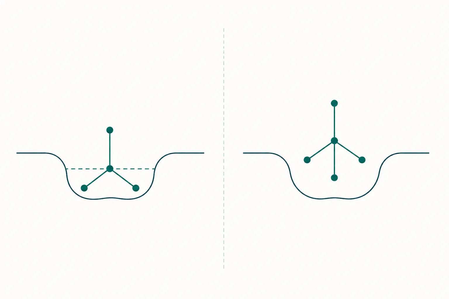
An alpha-carbon carrying four different substituents is a stereocentre, and it exists in two non-superimposable mirror-image forms designated L and D. The two forms have identical molecular formulae, identical masses, identical bond lengths and identical behaviour in any environment that is itself achiral. They differ only in the three-dimensional arrangement of the four groups around that single carbon. Nothing about that difference matters until the molecule meets something chiral, at which point it matters completely.
Proteolysis is exactly such an encounter. A serine protease does not simply approach a bond and break it. It must bind the substrate in an extended conformation across a groove, seat the side chain immediately preceding the scissile bond into a shaped subsite conventionally labelled S1, and hold the carbonyl carbon of that bond in precise register with a catalytic serine and the oxyanion hole that will stabilise the tetrahedral intermediate. Every element of that arrangement is geometric. Metalloproteases differ in catalytic chemistry but not in this requirement: the substrate must be presented in one specific orientation.
Inverting the stereocentre at that residue rotates the side chain out of its pocket and simultaneously reorients the backbone carbonyl. The enzyme can often still bind the peptide loosely, but it can no longer achieve the productive geometry, and catalysis does not proceed. The bond is not chemically stronger. It is simply no longer presentable. This is why the effect is local and positional rather than global, and why an unmodified cleavage site three residues away will still be cut at its normal rate.
The Single Substitution at the Cleavage Site
In practice, stabilising a peptide by this route begins with identifying where it is actually cut. Incubating the peptide with plasma, serum or a purified enzyme and following the fragments by liquid chromatography and mass spectrometry gives the cleavage map directly: the observed fragment masses locate the bonds being broken and the order in which they go. Only then is a substitution designed, at the residue immediately before or after the primary cut.
The historical examples are instructive because they are so economical. Vasopressin, a nine-residue hormone, has a circulating half-life of the order of ten to twenty minutes. Substituting D-arginine at position 8 and removing the amino terminus produced desmopressin, whose plasma half-life is measured in hours rather than minutes. The same edit also shifted receptor selectivity, which is the point that recurs throughout this subject: a change made for stability is never only a stability change 5.
Growth hormone secretagogue chemistry is dense with the same tactic. The short hexapeptide secretagogues carry D-tryptophan and D-phenylalanine at internal positions, and ipamorelin was built as a pentapeptide combining D-2-naphthylalanine and D-phenylalanine with alpha-aminoisobutyric acid at the amino terminus, giving a compound that was both stable and markedly more selective for growth hormone release than its predecessors in the rodent and porcine models used to characterise it 4. Somatostatin analogues follow the same pattern: the native fourteen-residue hormone survives one to three minutes, and analogues carrying D-phenylalanine and D-tryptophan survive orders of magnitude longer.
Retro-Inverso: Inverting the Whole Chain
The logical extreme of the single substitution is to invert every stereocentre in the molecule. Doing that alone gives the enantiomer, which is completely protease resistant and, against a chiral receptor, completely inactive. The retro-inverso strategy adds a second operation: reverse the sequence order as well. Reading the chain backwards while inverting every centre produces a molecule whose side chains occupy approximately the same positions in space as the parent, but whose backbone amide bonds all point the opposite way, with each carbonyl and amide nitrogen swapped 2.
The termini require attention, since reversal leaves an amine where a carboxyl belonged and vice versa; malonyl and gem-diamino residues are the conventional caps. The resulting molecule is invisible to proteases, because no natural enzyme is built to process a reversed backbone. Whether it retains activity depends entirely on how the parent binds. Where recognition depends on side chains presented from an extended segment, retro-inverso analogues can retain substantial affinity, which is why the approach has been most productive for linear epitopes and antibody-recognised sequences 2.
Where recognition depends on the backbone itself, it fails. Alpha-helices are stabilised by hydrogen bonds running in a defined direction along the chain, and a reversed backbone presents those donors and acceptors in the wrong orientation; beta-turns are similarly directional. This is the honest limitation of retro-inverso design, and it explains why, three decades after the concept was systematised, it remains a specialist technique rather than a general solution.
The Potency Trade-Off Is Not Automatic
The standard expectation is that stability is bought with affinity, because the receptor is as stereospecific as the protease. That expectation is correct often enough to be a useful default, and wrong often enough that it cannot be assumed. The outcome depends on local secondary structure. A D-residue is a helix breaker; substituting one into a helical binding segment usually destroys the fold and the activity together. In a loop, a turn or a terminal position, the same substitution is frequently neutral, and sometimes it stabilises the bound conformation and improves affinity.
The melanocortin field supplies the clearest positive case. Replacing methionine-4 with norleucine and phenylalanine-7 with D-phenylalanine in alpha-melanocyte-stimulating hormone produced an analogue that was substantially more potent than the native hormone and had a dramatically prolonged duration of action in frog and lizard skin bioassays, an effect attributed to both resistance to degradation and a more favourable bound conformation 3. That D-phenylalanine-7 substitution has been carried forward into essentially every synthetic melanocortin analogue studied since.
| Parent peptide | Substitution pattern | Effect on stability | Effect on activity |
|---|---|---|---|
| Vasopressin | D-arginine at position 8, deaminated terminus | Minutes to hours | Receptor selectivity shifted, not merely preserved |
| Alpha-melanocyte-stimulating hormone | D-phenylalanine at position 7 | Markedly prolonged in bioassay | Potency increased |
| Somatostatin | D-phenylalanine and D-tryptophan, within a cyclic scaffold | Minutes to hours | Subtype selectivity altered |
| Growth hormone secretagogue hexapeptides | D-tryptophan and D-phenylalanine at internal positions | Substantially increased | Retained, with improved selectivity in the pentapeptide |
| Membrane-active antimicrobial peptides | All residues inverted | Complete resistance | Fully retained |
That last row is the cleanest demonstration of the underlying principle. When an all-D enantiomer of a membrane-lytic peptide was synthesised and tested, it retained the antibacterial and channel-forming activity of the natural all-L form while becoming resistant to proteolysis, because the target, a lipid bilayer, has no chirality for the peptide to match 1. Stereospecificity is a property of the target, not of the peptide. Where the target is a folded protein, handedness is decisive; where it is a membrane, it is irrelevant.
How a D-Substitution Shows in Analysis
The analytical consequences follow directly from the chemistry, and they divide sharply into two cases. A peptide with one inverted residue among many L-residues is a diastereomer of the parent, not an enantiomer. Diastereomers have genuinely different shapes, different hydrophobic surface presentation and different chromatographic behaviour, so an epimer separates from its parent on an ordinary achiral reversed-phase column, typically as a resolved peak at a slightly shifted retention time. The masses are identical, so mass spectrometry alone will not distinguish them, and a peak eluting close to the main product with the correct mass is the classic signature of partial racemisation during synthesis.
That matters practically because solid-phase synthesis can racemise residues during activation, with cysteine and histidine the most vulnerable. The epimer is a real impurity with its own biological profile, and it is chromatographically visible only if the method was developed to resolve it. Purity reported from a gradient too steep to separate close-eluting diastereomers is purity reported against the wrong question.
The second case is harder. The full enantiomer of an all-L peptide has identical mass, identical achiral chromatographic retention and an identical ultraviolet spectrum. It cannot be distinguished by standard reversed-phase analysis at all. Three routes exist: circular dichroism, which returns a mirror-image spectrum and immediately reveals inverted handedness of the whole molecule; a chiral stationary phase; or complete acid hydrolysis followed by derivatisation of the released amino acids with a chiral reagent, which converts the enantiomeric pair into separable diastereomers and reports the D and L content of each residue. The last is the definitive method, and it is destructive, which is why it is a characterisation technique rather than a routine release assay 5.
References
- All-D amino acid-containing channel-forming antibiotic peptides
- A dozen years of retro-inverso peptidomimetics
- 4-Norleucine, 7-D-phenylalanine-alpha-melanocyte-stimulating hormone: a highly potent alpha-melanotropin with ultralong biological activity
- Ipamorelin, the first selective growth hormone secretagogue
- Therapeutic peptides: Historical perspectives, current development trends, and future directions