mechanisms
MOTS-c: A Peptide Written by the Mitochondrial Genome, and the AMPK Mechanism Proposed For It
MOTS-c is a 16-amino-acid peptide encoded inside the mitochondrial genome rather than the nuclear one. That single fact makes it a different kind of molecule from almost every other peptide discussed in this field, and it shapes the mechanism proposed for it.
MOTS-c is proposed to work by activating AMP-activated protein kinase, the cell's principal low-energy sensor, and it is proposed to do so indirectly: the peptide interferes with the folate–methionine cycle, the intermediate AICAR accumulates as a consequence, and accumulated AICAR activates AMPK 1. That is the mechanistic core, and it was described in the same 2015 report that identified the peptide. What makes MOTS-c unusual is not the pathway but the source. The peptide is encoded inside the mitochondrial genome — a 16-amino-acid product of a short open reading frame nested within the 12S ribosomal RNA gene — rather than in nuclear DNA, where essentially every other peptide discussed in this field originates 1. Almost everything demonstrated about its downstream effects has been demonstrated in mice or in cultured cells.
A peptide the nucleus did not write
The human mitochondrial genome is a circular molecule of 16,569 base pairs. The standard annotation gives it 37 genes: 13 protein-coding sequences, all of them subunits of the oxidative phosphorylation machinery, plus 22 transfer RNAs and 2 ribosomal RNAs. Everything else a mitochondrion needs — well over a thousand proteins — is encoded in the nucleus, translated in the cytoplasm and imported. That asymmetry has shaped how the organelle is described for decades: the nucleus instructs, the mitochondrion executes.
MOTS-c does not fit that description. Its reading frame sits inside MT-RNR1, the gene for the 12S ribosomal RNA, and it encodes a 16-residue peptide with no relationship to the electron transport chain 1. It is not the first such find. Humanin, a 24-residue peptide encoded within the 16S rRNA gene, was reported in 2001, and further small humanin-like peptides have been described from the same region. Reviews of this literature now treat mitochondrial-derived peptides as a category in their own right, with energy metabolism as the recurring theme across its members 4.
Why a 51-nucleotide reading frame went unnoticed
Two conventions in genome annotation kept sequences like this invisible. The first is a length cutoff. Automated open-reading-frame detection has historically discarded frames shorter than roughly 100 codons, on the reasonable statistical argument that short frames occur by chance in any sequence of sufficient length. A 51-nucleotide frame is filtered out before a human ever sees it. The second is nesting. The MOTS-c frame lies within an already-annotated gene, and an annotation pipeline that has assigned a stretch of sequence to a ribosomal RNA does not usually go looking for a protein inside it.
There is a third complication specific to mitochondria: the genetic code they use is not the standard one. In human mitochondria, AUA specifies methionine rather than isoleucine, UGA specifies tryptophan rather than a stop, and AGA and AGG act as stop codons rather than coding for arginine. The consequence is that the same run of nucleotides yields a different peptide, and different frame boundaries, depending on which code is applied to it. Where a given mitochondrial-derived peptide is actually translated — inside the organelle under the mitochondrial code, or in the cytoplasm under the standard one from an exported transcript — is a question the primary literature handles with more care than secondary summaries usually do.
The proposed mechanism: folate cycle, AICAR, AMPK
The 2015 report approached the mechanism through metabolomics rather than through a receptor hunt, and what it found was a shift in one-carbon metabolism. The folate–methionine cycle supplies one-carbon units for several biosynthetic routes, among them de novo purine synthesis. Purine synthesis requires 10-formyltetrahydrofolate at two separate formylation steps. MOTS-c was reported to restrict flux through this cycle, and the reported downstream consequence was accumulation of AICAR, the purine intermediate immediately upstream of the second of those formylation steps 1.
That accumulation is the pivot of the whole model, because AICAR in its phosphorylated form is a structural mimic of AMP. AMPK is a heterotrimeric kinase whose gamma subunit carries nucleotide-binding sites; occupancy of those sites by AMP, or by an AMP-like molecule, promotes the phosphorylation state that renders the kinase active. A metabolic block that raises an AMP mimic therefore switches on the low-energy sensor without the cell's actual ATP having fallen. Reviews of mitochondrial-derived peptides place this AMPK dependence at the centre of the class's reported metabolic effects 4.
Activated AMPK does broadly what its role as an energy sensor implies. It promotes catabolic, ATP-generating processes — glucose uptake, fatty acid oxidation — and restrains anabolic, ATP-consuming ones, including lipid synthesis and protein synthesis via mTORC1. Read against that, the metabolic phenotype reported in mice given MOTS-c is what an AMPK-activating agent would be predicted to produce: resistance to diet-induced obesity and improved insulin sensitivity in animals fed a high-fat diet 1. The internal consistency of the mechanism and the phenotype is a point in the model's favour. It is not a substitute for testing the phenotype in another species.
Retrograde signalling and nuclear translocation
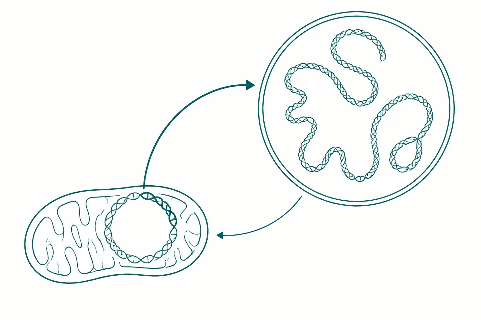
Communication between the nucleus and the mitochondrion runs in both directions, and the two directions have names. Anterograde signalling is the familiar one: the nucleus regulates mitochondrial biogenesis, composition and turnover. Retrograde signalling runs the other way — the organelle reports its state to the nucleus and the nucleus adjusts transcription in response. Retrograde signals were classically described as metabolites and second messengers: calcium, reactive oxygen species, shifts in the NAD ratio. A peptide encoded by the mitochondrial genome would be a different sort of retrograde signal entirely, because the message would carry mitochondrial genetic information rather than merely a metabolic readout.
That is the claim made in 2018. Under metabolic stress — glucose restriction, serum deprivation, oxidative stress — MOTS-c was reported to move from the cytoplasm into the nucleus, in a manner dependent on AMPK, and there to associate with stress-responsive transcription factors and regulate a set of nuclear genes, including genes carrying antioxidant response elements 2. The peptide was described not as a hormone acting on a surface receptor but as a regulator operating at the level of transcription in the compartment that would ordinarily be issuing the instructions.
The model system matters here and is easy to lose sight of. This work was done in cultured cells and in mouse tissue. It establishes that the translocation occurs and is stress-dependent in those systems. It does not establish what fraction of endogenous MOTS-c in an intact human ever reaches a nucleus, nor whether nuclear localisation contributes measurably to any physiological outcome in a person. The concept is well posed and the cellular evidence is real; the leap from there to human physiology has not been made.
The exercise connection
The 2021 work is where a human measurement enters the record, and it is worth being exact about what kind of measurement it is. MOTS-c was reported to be exercise-responsive: levels rose in skeletal muscle and in the circulation following exercise, in mice and also in human participants, with the muscle response substantially larger than the plasma response 3. This is observational. Blood and tissue were sampled around exercise and an endogenous peptide was quantified. Nothing was administered to the human participants, and no clinical outcome was assigned.
The interventional half of the same paper is entirely animal work. MOTS-c administration in mice was reported to improve physical performance across young, middle-aged and old cohorts, with effects on running capacity and on markers of muscle homeostasis, and the paper framed the peptide as a regulator of age-dependent physical decline 3. Those two halves are frequently reported together as though they were one finding. They are not. The human half says a peptide changes with exercise. The mouse half says giving the peptide changes performance. Neither implies the other, and the combination is not evidence that giving the peptide to a person would do anything.
Model systems, stated plainly
| Reported finding | Model system | What it establishes |
|---|---|---|
| 16-residue peptide encoded within the mitochondrial 12S rRNA gene | Human mitochondrial genome sequence; cultured cells | Sequence-level; well supported |
| Restriction of folate-cycle flux and accumulation of AICAR | Metabolomics in cultured cells and mouse tissue | Proposed mechanism, biochemically coherent |
| AMPK activation downstream of AICAR accumulation | Cultured cells; mouse tissue | Proposed mechanism |
| Resistance to diet-induced obesity; improved insulin sensitivity | Mice on a high-fat diet | Animal only |
| Nuclear translocation under metabolic stress | Cultured cells; mouse tissue | Cellular; not shown to drive a human outcome |
| Regulation of nuclear stress-response gene expression | Cultured cells | In vitro |
| Rise in muscle and circulating levels after exercise | Mice; human observational sampling | Association in humans; no causal claim |
| Improved physical capacity in aged animals | Mice, including old cohorts | Animal only |
| Effect of administration in humans | No published controlled trial | Not established |
Read down the third column and the shape of the evidence is clear. The molecular and cellular claims are the strongest part of the record, and they are genuinely interesting independent of any therapeutic question — a peptide product of the mitochondrial genome that acts on nuclear transcription is a real addition to cell biology. The whole-organism metabolic claims sit one species removed from humans. The human column contains one thing: a measurement of an endogenous peptide that moves with exercise.
What the evidence does not establish
- No controlled human trial of MOTS-c administration has been published. There is no human efficacy result for any endpoint, and no human safety dataset.
- No human pharmacokinetic profile is published. Absorption, distribution, half-life and clearance of administered MOTS-c in humans are unknown.
- Exercise-responsiveness in humans is a correlation. A peptide rising alongside exercise may be a mediator of adaptation, a marker of it, or a byproduct; the observational design cannot distinguish these.
- The mouse obesity and insulin-sensitivity results were generated under a high-fat-diet challenge. Effects observed against an induced metabolic insult in a rodent do not transfer automatically to unstressed physiology or to another species.
- AMPK activation is not a uniformly desirable end state. AMPK restrains anabolic signalling, including mTORC1, and chronic pharmacological activation of a master metabolic switch has consequences that short experiments are not designed to detect.
- The relationship between endogenous MOTS-c concentrations and any administered quantity is unquantified in humans, so there is no basis for reasoning about physiological relevance.
- Material sold under the name is not covered by any pharmacopoeial standard, so identity and purity are supplier-dependent rather than assured.
How to read this literature
Three habits keep this field legible. First, check whether a stated result concerns endogenous MOTS-c or administered MOTS-c; those are different experiments answering different questions, and the exercise literature contains both 3. Second, check the species before accepting a metabolic claim, because the obesity and insulin-resistance findings are mouse findings and are usually reported without that qualifier 1. Third, treat the mechanism and the outcome as separate propositions: the folate-cycle-to-AICAR-to-AMPK route can be well evidenced at the level of cell biochemistry while telling you nothing about whether administering the peptide produces an outcome in a person 4.
The discovery itself deserves the attention it has received. A short open reading frame hidden inside a ribosomal RNA gene, in a genome that was considered fully catalogued, producing a peptide that appears to signal back to the nucleus under stress, is a substantive revision to how the organelle is understood 2. The overstatement does not usually happen at that stage. It happens when the novelty of the biology is transferred to a claim about what the compound does when given to a human — a claim on which the published literature is, at present, silent.
References
- The mitochondrial-derived peptide MOTS-c promotes metabolic homeostasis and reduces obesity and insulin resistance
- The Mitochondrial-Encoded Peptide MOTS-c Translocates to the Nucleus to Regulate Nuclear Gene Expression in Response to Metabolic Stress
- MOTS-c is an exercise-induced mitochondrial-encoded regulator of age-dependent physical decline and muscle homeostasis
- Mitochondrial-derived peptides in energy metabolism