fundamentals
L-Carnitine Is Not a Peptide: What It Does and What the Evidence Supports
Carnitine is a single small molecule of about 161 daltons that sits in peptide catalogues for commercial reasons rather than chemical ones. Its role in fatty acid transport is textbook biochemistry; the fat-loss claim built on top of that role is a separate question with a much weaker answer.
No on both counts: L-carnitine is not a peptide, and in people who are not carnitine-deficient there is no strong evidence that taking it produces meaningful fat loss. It is a quaternary ammonium compound with a molecular weight of about 161 daltons — a single small molecule, not a chain of amino acids joined by peptide bonds. It appears in peptide catalogues for commercial reasons rather than chemical ones. Its actual biochemistry is not in dispute and is genuinely important: without carnitine, long-chain fatty acids cannot cross into the mitochondrial matrix to be oxidised 1. What does not follow from that fact is the claim most often built on top of it.
A single molecule, not a chain
Carnitine is 3-hydroxy-4-(trimethylammonio)butanoate. The trimethylammonio group is a permanently charged quaternary nitrogen, which is what makes the molecule a zwitterion at physiological pH and keeps it from crossing membranes freely. There is one chiral centre, and only the L enantiomer is biologically active; D-carnitine is not merely inert but competes with the active form at the same transporters.
The confusion with peptides has a plausible-sounding root. Carnitine is derived from amino acids: it is synthesised in the liver, kidney and brain from a lysine residue trimethylated on a protein, using methionine as the methyl donor, then released by proteolysis and carried through four enzymatic steps to the finished molecule, with ascorbate, iron, pyridoxine and niacin required as cofactors 1. So amino acids are the raw material. That is a biosynthetic relationship, not a structural one. Cholesterol is made from acetyl-CoA and nobody calls it a carbohydrate.
The structural test is straightforward and carnitine fails it. A peptide is a polymer: two or more amino acid residues joined by amide bonds, with a defined N-terminus, a C-terminus and a repeating backbone. Carnitine is a seven-carbon monomer. There is nothing to sequence, no direction to read, no residue count.
| Feature | L-carnitine | A peptide |
|---|---|---|
| Class | Quaternary ammonium compound | Amino acid polymer |
| Molecular weight | About 161 Da | Roughly 300 Da upward |
| Peptide bonds | None | One or more, by definition |
| Sequence | Not applicable | Defined, read N-terminus to C-terminus |
| Origin | Enzymatic synthesis from a trimethylated lysine residue | Ribosomal translation or chemical synthesis |
| Digestive fate | Absorbed intact; not cleaved by proteases | Susceptible to proteolysis |
What carnitine actually does
The function is well established and worth stating precisely, because the precision is where the argument turns. Long-chain fatty acids are activated in the cytosol to acyl-CoA esters. Acyl-CoA cannot cross the inner mitochondrial membrane, and beta-oxidation happens in the matrix. Carnitine solves that transport problem.
Carnitine palmitoyltransferase I, on the outer mitochondrial membrane, transfers the acyl group from CoA to carnitine, producing acylcarnitine. A carnitine-acylcarnitine translocase in the inner membrane exchanges that acylcarnitine inward for a free carnitine moving outward. On the matrix side, carnitine palmitoyltransferase II transfers the acyl group back onto CoA, regenerating acyl-CoA inside the mitochondrion and releasing free carnitine to make the return trip. Beta-oxidation then proceeds on the acyl-CoA.
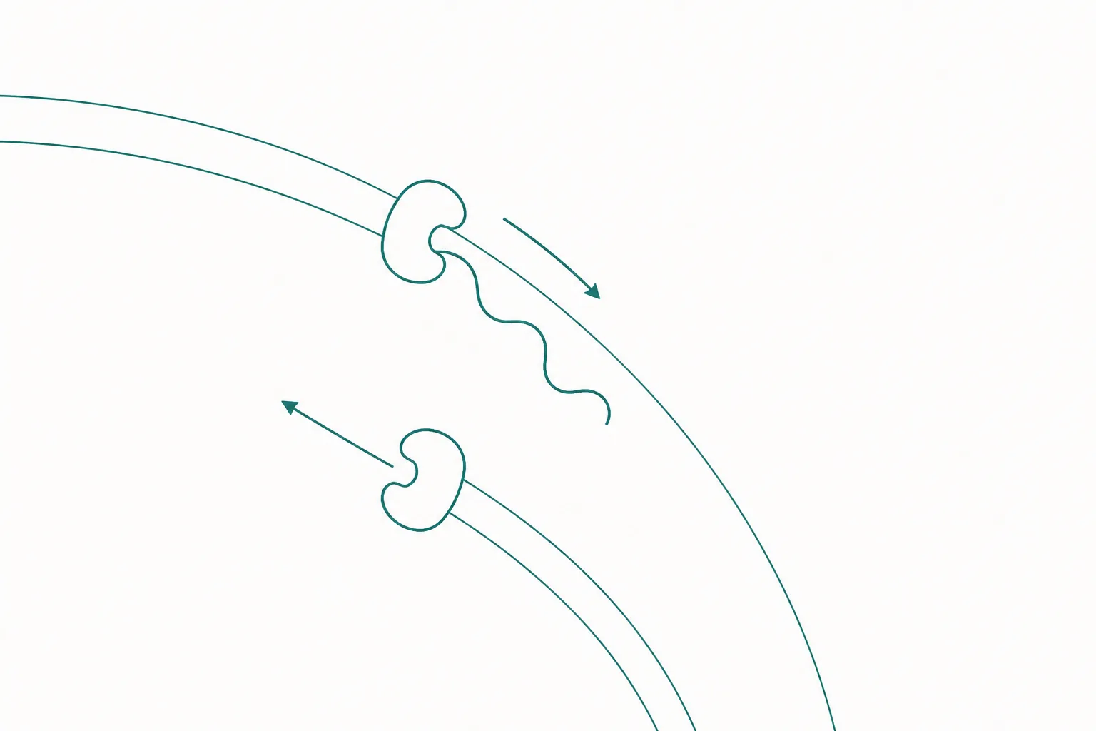
Nothing here is hypothetical. It is standard metabolic biochemistry, and losing it has severe, well-characterised consequences: long-chain fatty acids cannot be oxidised, and the tissues most dependent on fat as fuel — cardiac and skeletal muscle — fail first 1. Around 95% of body carnitine sits in skeletal muscle.
Required is not the same as rate-limiting
This is the most useful idea here, and it generalises far beyond carnitine. A required cofactor increases flux through a pathway only if it is the thing currently constraining flux. If the limit lies elsewhere — substrate availability, enzyme activity, oxygen delivery, ADP demand — adding more cofactor changes nothing, because it was never the bottleneck.
The commercial argument runs: fat oxidation requires carnitine, therefore more carnitine means more fat oxidation. The first clause is true. The second does not follow. In a healthy adult with ordinary dietary intake and intact synthesis, skeletal muscle carnitine sits well in excess of what the transferase reaction requires under most conditions. Control of the shuttle is exerted elsewhere — principally at carnitine palmitoyltransferase I, which is inhibited by malonyl-CoA, the molecule signalling that the cell is in a fed, lipogenic state. That inhibition is the switch. Adding carrier molecules does not release a brake applied somewhere else.
The exception shows the structure of the argument. Where carnitine genuinely is limiting — because a transporter defect, a metabolic disorder or a drug has depleted it — restoring it restores flux, and the clinical effect can be dramatic. The absence of any comparable effect in replete individuals is evidence about which regime they are in, not about whether the biochemistry is real.
Why muscle carnitine is hard to raise
There is a second obstacle, independent of the first. Even if muscle carnitine were limiting, raising it by mouth is difficult. Skeletal muscle concentrates carnitine to roughly a hundred times the plasma level through an active, sodium-dependent transporter — OCTN2, the product of the SLC22A5 gene. Transport against a steep gradient is saturable, and at ordinary plasma concentrations the transporter already operates near capacity.
The consequence is a decoupling between the two compartments. Oral intake raises plasma carnitine readily; the kidney increases excretion, and muscle content moves very little. Human studies that have meaningfully raised muscle carnitine have generally had to force the issue by manipulating insulin, which stimulates OCTN2-mediated uptake, and have required sustained administration over months. Whole-body homeostasis is defended by efficient renal reabsorption, which is why vegetarians, taking in far less from diet, maintain functional tissue stores despite lower plasma levels 1.
Where supplementation genuinely matters
The legitimate clinical picture has a familiar shape: replacement in a defined deficiency state is a different proposition from supplementation in a replete one, in the same way that growth hormone replacement in documented deficiency differs from growth hormone taken by someone who has plenty. The settings in which carnitine repletion is established are specific and diagnosable.
- Primary systemic carnitine deficiency, caused by loss-of-function mutations in SLC22A5. Cellular uptake and renal reabsorption both fail; presentation includes cardiomyopathy, skeletal myopathy and hypoketotic hypoglycaemia. Repletion is disease-modifying.
- Secondary deficiency from inborn errors of metabolism, particularly organic acidaemias, in which accumulating acyl groups are conjugated to carnitine and excreted as acylcarnitines, depleting the free pool.
- Secondary deficiency from drug therapy, most characteristically long-term valproate, which drives the same acylcarnitine-mediated depletion.
- Haemodialysis. Carnitine is small, water-soluble and poorly protein-bound, so dialysis removes it efficiently; long-term patients can become depleted, and repletion is recognised in that population.
- Certain fatty acid oxidation disorders, where the intervention depends on which enzyme is affected, because supplying carnitine to a blocked pathway is not always benign.
In each case the reasoning is the same, and it is the reasoning that fails for the general population: the carrier is demonstrably depleted, so supplying it restores a function that was actually lost.
The exercise and body-composition evidence
Reported honestly, the human evidence is modest and mixed, and it does not point where the marketing points. Trials in healthy and trained adults have looked for increased fat oxidation, reduced fat mass and improved endurance performance. Results are inconsistent and effect sizes small where they appear at all; studies that measured muscle carnitine alongside the outcome frequently found muscle content unchanged, which makes any mechanistic interpretation difficult.
The more interesting signal sits in a different outcome. A body of work summarised in a 2018 review in Nutrients concerns recovery rather than fat loss: markers of exercise-induced muscle damage, perceived soreness and indices of oxidative stress after damaging exercise, with proposed mechanisms involving blood flow and tissue hypoxia rather than substrate oxidation 4. The trials are mostly small, heterogeneous in protocol and population, and often rest on inherently subjective soreness scales. It is a reasonable hypothesis with supporting data, not an established effect.
The asymmetry is worth noting. The claim that sells the product — increased fat oxidation leading to fat loss — has the weakest support. The claim with the most plausible supporting data concerns post-exercise recovery, and is rarely the one advertised.
The TMAO finding, and what it does and does not show
In 2013 a group publishing in Nature Medicine reported that intestinal microbiota metabolise dietary carnitine to trimethylamine, which is absorbed and oxidised by hepatic flavin-containing monooxygenases to trimethylamine N-oxide, and that TMAO is associated with atherosclerosis 2. The paper is important and deserves describing accurately rather than being either dismissed or inflated.
The mechanistic component is substantially murine. In mice, dietary carnitine accelerated atherosclerosis in an atherogenic background strain; suppressing the gut microbiota with antibiotics abolished both TMAO production and the acceleration; and germ-free animals did not produce TMAO from carnitine until colonised. That is a coherent causal chain, established in a mouse model of a disease mice do not develop the way people do.
The human component is associative. Plasma TMAO was associated with prevalent cardiovascular disease and with incident events in a large clinical cohort. Association does not establish causation, and TMAO plausibly marks several things at once: diet composition, renal function — it is renally cleared, so impaired kidneys raise it — and microbiota composition. Whether it causally contributes to cardiovascular events in humans, or mainly reports other risk, is still debated.
One further detail from the same work matters for interpretation. Production is not a fixed property of the amount ingested; it is a property of the gut community. Habitual omnivores produced substantially more TMAO after a carnitine challenge than vegans and vegetarians did, because chronic dietary exposure selects for the bacterial taxa performing the conversion 2. The same intake in two people can therefore yield very different exposure. This is a real and interesting signal about a diet-microbiome-host axis. It is not a settled harm.
The diabetes literature
Carnitine is an active research interest in type 2 diabetes, and the interest should be reported at the size it actually is. Incomplete fatty acid oxidation in skeletal muscle generates acyl intermediates implicated in insulin resistance, and carnitine buffers them by conjugating them into exportable acylcarnitines; plasma acylcarnitine profiles are consequently studied as readouts of mitochondrial fuel handling. A 2018 review in Nutrition & Diabetes surveys this ground alongside trial work on the acetylated and propionylated derivatives in glycaemic control and diabetic neuropathy 3. The mechanism is reasonable; it is not a demonstrated therapy, and it says nothing about body composition in people without metabolic disease.
Why the category error is worth correcting
Filing carnitine among peptides is not a harmless shelving convention. It borrows credibility across a category line. The word peptide signals research-grade, mechanistically specific, biologically sophisticated, and a compound placed on that shelf inherits the impression without earning it, while anyone reasoning about it applies expectations drawn from an entirely different chemistry.
The correction is small and clarifying. Carnitine is a well-understood small molecule with a well-understood job, a defined set of conditions in which supplying it is genuinely useful, a weak evidence base for the use that drives most consumer sales, and one unresolved question about its microbial metabolism.
The transferable lesson is the one in the middle of this article. Whenever a compound is promoted on the grounds that a process requires it, the question is not whether the requirement is real. It usually is. The question is whether it is currently unmet.