immune and mitochondrial peptides
LL-37: The Human Cathelicidin
LL-37 is the only cathelicidin humans make. It kills bacteria by physically disrupting their membranes, signals to immune cells through a formyl peptide receptor, and is transcriptionally controlled by vitamin D. It is also a case study in why antimicrobial peptides keep failing in clinical development.
Almost every multicellular organism makes short peptides that kill microbes on contact. Frogs make magainins, insects make cecropins, cattle make more than a dozen distinct cathelicidins. Humans make one. It is called LL-37, it is 37 residues long, it begins with two leucines, and it is the terminal product of a single gene on chromosome 3. It is one of the best-characterised molecules in innate immunity and one of the least successful in drug development, and those two facts are connected.
Host defence peptides, and where LL-37 comes from
The class was named twice. Antimicrobial peptide describes what these molecules do in a test tube. Host defence peptide describes what they appear to do in an organism, which is a broader job that includes recruiting cells, neutralising bacterial products and shaping wound repair. Several thousand have been catalogued across the animal, plant and bacterial kingdoms. They share almost no sequence homology, and they converge instead on physical properties: short length, net positive charge, and the ability to fold into a structure that separates charged residues from hydrophobic ones 1.
In humans the two main families are the defensins, which are beta-sheet peptides stabilised by three disulphide bonds, and the cathelicidins, which are defined not by their mature sequence but by their precursor. Every cathelicidin is translated as a two-part protein: a highly conserved N-terminal domain called cathelin, structurally related to the cathepsin L inhibitors, and a C-terminal antimicrobial domain that varies enormously between species.
The human gene, CAMP, was described in 1996 and its product initially called FALL-39 after its first four residues and its predicted length 2. The precursor protein, hCAP18, is stored in the specific granules of neutrophils and in the secretory vesicles of epithelial cells. It is inert while intact. Cleavage by proteinase 3 releases the mature 37-residue peptide, and only then does antimicrobial activity appear. In skin the processing is different again: kallikrein-related peptidases 5 and 7 cut hCAP18 to a set of shorter fragments with distinct activity profiles, so the peptide present in sweat and epidermis is not necessarily the same species that neutrophils release.
That storage-as-precursor arrangement matters. A peptide that lyses membranes cannot safely be held in an active form inside a cell that has membranes of its own. The cathelin domain is a safety catch, and the location of the protease determines which mature product appears where.
The amphipathic helix and what it does to a membrane
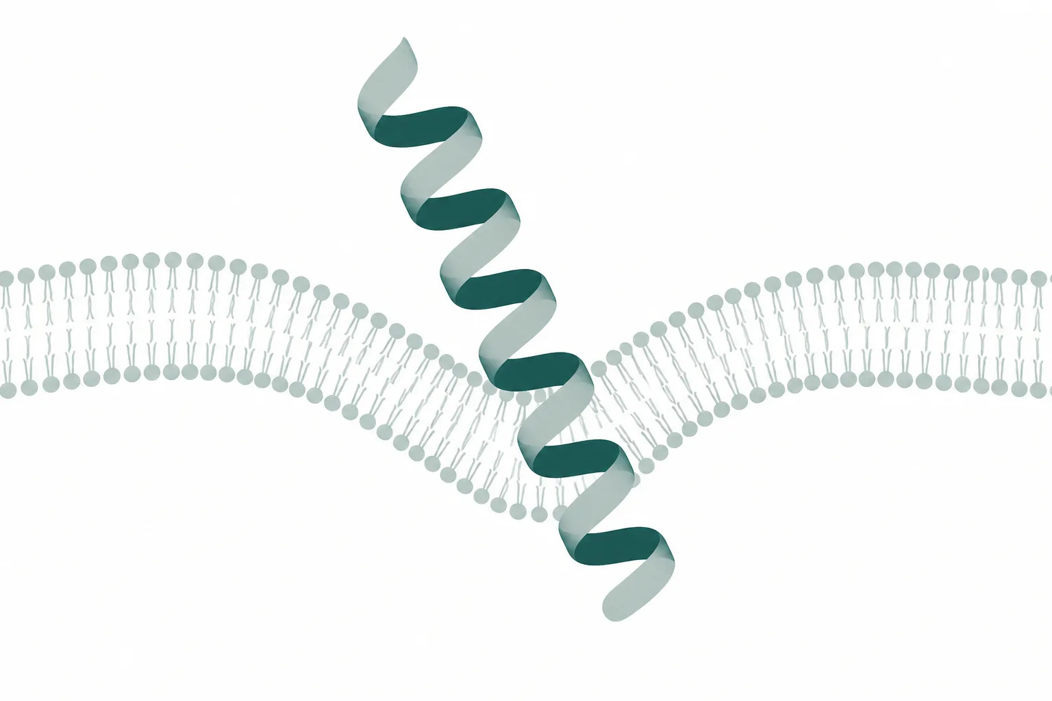
In water LL-37 is largely disordered. On contact with an anionic surface it folds into an alpha helix, and the helix is amphipathic: because a helical turn is roughly 3.6 residues, the peptide sequence is arranged so that lysine and arginine residues cluster on one face while leucine, phenylalanine and isoleucine cluster on the other. The peptide carries a net charge near plus six at physiological pH. Charge draws it to the membrane. Hydrophobicity drives it in.
Selectivity for microbes over host cells is electrostatic, not molecular recognition. Bacterial surfaces are strongly anionic: Gram-negative outer membranes present lipopolysaccharide, Gram-positive walls present teichoic acids, and the inner leaflet lipids include phosphatidylglycerol and cardiolipin. The outer leaflet of a mammalian plasma membrane is dominated by zwitterionic phosphatidylcholine and sphingomyelin, and it contains cholesterol, which stiffens the bilayer and resists peptide insertion. The peptide is not identifying bacteria. It is following charge 1.
What happens after insertion is described by several competing models. In the barrel-stave model peptides assemble into a defined transmembrane pore. In the toroidal pore model they bend the bilayer so that lipid head groups line the channel with them. In the carpet model they accumulate on the surface until the membrane disintegrates detergent-like. LL-37 is usually assigned to the carpet or toroidal descriptions, and the honest position is that the mechanism is concentration-dependent and probably not a single event.
The second job: receptor signalling and immune modulation
The concentrations required to kill bacteria in a buffer are considerably higher than those found in most tissues. That gap prompted a reassessment of what the peptide is actually for, and the answer that emerged is that direct killing may be secondary to signalling.
In 2000 LL-37 was shown to chemoattract human neutrophils, monocytes and T cells through formyl peptide receptor-like 1, now normally called FPR2, a G protein-coupled receptor previously known for responding to bacterial formylated peptides 3. This is receptor-mediated activity at nanomolar concentrations, orders of magnitude below the lytic range. The same peptide therefore has two mechanisms operating on entirely different concentration scales, and confusing them is a common error when reading this literature.
Other described activities include binding and neutralising lipopolysaccharide, which blunts the septic response to Gram-negative debris; transactivation of the epidermal growth factor receptor in keratinocytes, linked to re-epithelialisation of wounds; promotion of angiogenesis; and modulation of neutrophil apoptosis. The peptide also has a documented pathological side. In psoriatic skin, LL-37 forms complexes with self-DNA that are taken up by plasmacytoid dendritic cells and activate TLR9, converting a molecule that normally ignores host nucleic acid into a driver of chronic inflammation. A host defence peptide is not automatically a beneficial one.
Vitamin D controls the gene
The CAMP promoter contains a vitamin D response element, which places cathelicidin transcription directly downstream of vitamin D receptor activation. The mechanistic demonstration came in 2006. Engagement of TLR2 on human monocytes upregulated both the vitamin D receptor and CYP27B1, the enzyme that converts circulating 25-hydroxyvitamin D into the active 1,25-dihydroxy form. The cell therefore generates its own active vitamin D locally, in response to a bacterial signal, and uses it to switch on cathelicidin 4.
The consequence reported in that work is the part worth retaining: serum from donors with low 25-hydroxyvitamin D was less able to support this induction than serum from replete donors, and supplementing the deficient serum restored it 4. This gave a concrete molecular route from vitamin D status to antibacterial capacity, and it is the strongest mechanistic link between the two that exists.
Two caveats belong with it. First, a mechanism that operates in cultured human monocytes does not establish that vitamin D supplementation reduces infection rates in a population, and large supplementation trials with infection endpoints have produced mixed results. Second, the vitamin D response element in the cathelicidin promoter arose from a primate-specific transposable element insertion and is not present in rodents. Mouse cathelicidin is not vitamin D regulated. Any mouse experiment testing this axis is testing something the mouse does not have.
Why antimicrobial peptides keep failing in development
The class has been in development for more than thirty years. Its promise was obvious from the start: broad spectrum, fast killing, a physical mechanism resistant to the usual resistance routes, and a natural template in every organism examined. The record of approvals does not match that promise, and the reasons are consistent enough to be worth listing rather than treating case by case 5.
| Candidate | Derived from | Route and target | Outcome |
|---|---|---|---|
| Pexiganan | Magainin, frog skin | Topical, diabetic foot infection | Failed to gain approval across two separate submissions |
| Iseganan | Protegrin, porcine | Oral rinse, mucositis | Phase 3 failed to beat placebo; a separate pneumonia study was halted |
| Omiganan | Indolicidin, bovine | Topical, catheter site infection | Missed its primary endpoint in phase 3 |
Every entry in that table is topical, and that is the first part of the explanation. Systemic development is blocked before efficacy can even be assessed. A cationic amphipathic peptide that lyses anionic membranes will lyse red blood cells at high enough concentration, and the therapeutic window between antibacterial and haemolytic activity is narrow. Given intravenously, these molecules also bind serum albumin and lipoproteins, which sequesters them, and are cleared by proteases within minutes.
The second part is salt. The activity measured in a low-ionic-strength buffer is not the activity present in tissue. Screening assays are commonly run in dilute media because that is where the peptides perform, and raising sodium chloride to physiological concentration can raise minimum inhibitory concentrations by an order of magnitude or more. Divalent cations and serum proteins compound the problem. A large fraction of the published potency data for this class was generated under conditions that do not correspond to the environment in which a drug would have to work 5.
The remaining obstacles are practical rather than biological. Solid-phase synthesis of a 37-residue peptide at tonne scale is expensive relative to a small molecule. Peptides raise immunogenicity questions that small molecules do not. And there is a specific concern unique to this class: an antibiotic that mimics an endogenous host defence peptide risks selecting for organisms cross-resistant to the innate immune system itself, which is a category of harm conventional antibiotics do not carry.
That reframing is where the field has landed. If the peptide is a signalling molecule that also happens to kill bacteria under favourable ionic conditions, then developing it as a replacement antibiotic was always fighting its pharmacology. Programmes targeting the receptor-mediated arm, at nanomolar rather than micromolar concentrations, are working with the molecule rather than against it. Whether that arm produces an approved medicine remains unresolved, and thirty years of history suggest reserving judgement.
References
- Antimicrobial peptides of multicellular organisms
- The human gene FALL39 and processing of the cathelin precursor to the antibacterial peptide LL-37 in granulocytes
- LL-37, the neutrophil granule- and epithelial cell-derived cathelicidin, utilizes formyl peptide receptor-like 1 (FPRL1) as a receptor to chemoattract human peripheral blood neutrophils, monocytes, and T cells
- Toll-like receptor triggering of a vitamin D-mediated human antimicrobial response
- Designing antimicrobial peptides: form follows function