immune and mitochondrial peptides
Humanin and the Mitochondrial-Derived Peptide Family
Humanin was found by accident in a screen of brain tissue from an Alzheimer's patient, and it turned out to be encoded inside the mitochondrial genome. It opened a family of short peptides read from mitochondrial DNA, and almost every quantitative claim about them rests on assays that are difficult to trust.
The human mitochondrial genome is 16,569 base pairs of circular DNA and was thought to be fully catalogued decades ago: 13 protein-coding genes, 22 transfer RNAs, 2 ribosomal RNAs, all of them components of the respiratory chain or the machinery that builds them. Nothing in it looked like a signalling molecule. Then a screen looking for something else entirely turned up a peptide that protected neurons from death, and its sequence mapped to a short stretch inside one of the ribosomal RNA genes. That peptide is humanin, and it opened a category that the annotation had missed.
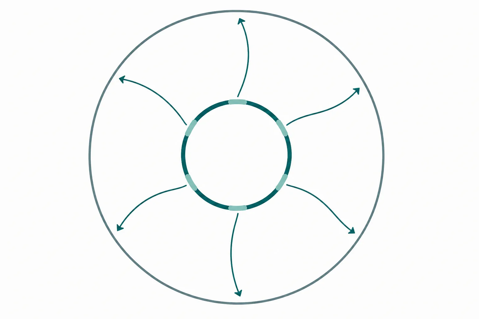
Short reading frames in a genome nobody expected to encode peptides
Genome annotation has a minimum length threshold. Open reading frames below roughly 100 codons were routinely discarded as noise, because in a genome of any size short reading frames occur by chance in large numbers and screening them all was impractical. That convention was applied to the mitochondrial genome as it was to the nuclear one, and it is the reason a set of functional 20-residue peptides sat unannotated inside genes that had been sequenced in 1981.
The mitochondrial genome makes the problem worse in a specific way, because it does not use the standard genetic code. In human mitochondria, ATA specifies methionine rather than isoleucine, TGA specifies tryptophan rather than a stop, and AGA and AGG function as stop codons rather than arginine. A reading frame therefore looks different depending on which code is applied, and a sequence that terminates early under the standard code may run on under the mitochondrial one. The 21-residue and 24-residue forms of humanin are exactly this: the same DNA read under two different codes in two different compartments.
There is a second complication that has never been fully resolved. If these peptides are translated inside the mitochondrion by the mitoribosome, they must then leave it to reach the cytosol, the nucleus or the circulation, and no export machinery for mitochondrial translation products has been described. The alternative is that mitochondrial transcripts reach the cytoplasm and are translated there by cytoplasmic ribosomes, which would make the name mitochondrial-derived accurate as to genomic origin but misleading as to site of synthesis. Both routes have supporters and the question is open.
How humanin was found
The 2001 discovery was not a search for mitochondrial peptides. It was a search for anything that could stop neurons dying. The investigators built a complementary DNA library from the occipital lobe of a patient who had died with Alzheimer's disease, choosing that region specifically because it is comparatively spared while the hippocampus and association cortex are destroyed. The reasoning was that surviving tissue might be expressing something protective.
The library was screened in a neuronal cell system for clones that rescued cells from death induced by mutant familial Alzheimer's disease genes and by amyloid beta. One clone did so across a wide range of such insults, which was itself notable because those insults act through different upstream routes. The active element was a short peptide, and its sequence corresponded to a region of the mitochondrial 16S ribosomal RNA gene 1. The investigators named it humanin.
Mechanistic work followed quickly along two lines. In 2003 humanin was reported to bind Bax, the pro-apoptotic protein whose translocation from cytosol to mitochondrial outer membrane commits a cell to intrinsic apoptosis, and to prevent that translocation 2. Separately, humanin was found to bind insulin-like growth factor-binding protein 3, placing it in contact with a growth factor axis that regulates survival and apoptosis in its own right 3. Later work added extracellular receptors, including formyl peptide receptor 2 and a trimeric receptor complex comprising CNTFR, WSX-1 and gp130 that signals through STAT3, which would make humanin a peptide acting both inside the cell and on its surface.
What the family now contains
Humanin remained an isolated curiosity for a decade. The category formed in 2015, when MOTS-c was described: a 16-residue peptide read from a short open reading frame in MT-RNR1, the 12S ribosomal RNA gene, reported to influence the folate one-carbon cycle, to be associated with AMP-activated protein kinase activation, and to reduce diet-induced insulin resistance in mice 4. A second peptide from a second mitochondrial gene made the first one look like a class rather than an exception.
The following year six further peptides were reported from alternative reading frames within MT-RNR2 and named small humanin-like peptides, SHLP-1 through SHLP-6 5. They are not fragments of humanin. They are separate reading frames within the same gene, and the reported activities differ substantially between them, with SHLP-2 and SHLP-3 attracting most attention for effects on apoptosis and metabolism while others showed little activity in the same assays.
| Peptide | Mitochondrial gene | Length | Principal reported activity |
|---|---|---|---|
| Humanin | MT-RNR2 (16S rRNA) | 21 or 24 residues by compartment | Cytoprotection, Bax interference, receptor signalling |
| MOTS-c | MT-RNR1 (12S rRNA) | 16 residues | Metabolic regulation, folate cycle, nuclear translocation |
| SHLP-1 to SHLP-6 | MT-RNR2, alternative frames | Around 20 to 38 residues | Variable; apoptosis and metabolism for some members |
A structural observation is worth making about that table. Every confirmed member is read from a ribosomal RNA gene. That is not obviously meaningful, and it may simply reflect where investigators have looked, since the two ribosomal RNA genes are the longest non-protein-coding stretches in the mitochondrial genome and therefore the most promising places to search for unannotated frames. Whether the protein-coding genes also harbour such frames has not been systematically resolved.
Retrograde signalling
The conceptual interest of this family is direction of information flow. The nucleus controls the mitochondrion comprehensively: the overwhelming majority of the roughly 1,100 proteins in a mitochondrion are nuclear-encoded, translated in the cytosol and imported. Communication in the other direction, mitochondrion to nucleus, is called retrograde signalling, and until recently it was understood entirely in terms of indirect messengers. Falling ATP, rising reactive oxygen species, altered calcium handling, changes in the ratio of oxidised to reduced nicotinamide adenine dinucleotide: the nucleus infers mitochondrial status from these metabolic consequences.
A peptide encoded by mitochondrial DNA that acts on nuclear processes would be a different kind of channel entirely. It would carry a specific message rather than a diffuse metabolic signal, and the message would be encoded in the organelle's own genome. MOTS-c has been reported to translocate to the nucleus under metabolic stress and to associate with stress-responsive transcription factors, which is the most direct evidence for such a channel 4. Humanin, by contrast, is largely described as acting through cell-surface receptors, which would make it an endocrine or paracrine signal rather than an intracellular one.
If this is real at physiological concentrations, it has an evolutionary implication. The mitochondrial genome would not be merely a vestigial remnant retained because a handful of hydrophobic respiratory subunits are difficult to import. It would be an active regulatory element with its own outward signalling capacity. That is a substantial claim, and it should be weighed against the measurement problems described in the next section rather than separately from them.
Why quantifying these peptides is so difficult
A large fraction of what is claimed about this family is quantitative: that circulating concentrations fall with age, that they rise with exercise, that they differ between disease states. Those claims depend entirely on the assays used to generate them, and the assays are the weakest part of the field.
- Reported circulating concentrations of humanin span several orders of magnitude across published studies. That spread cannot represent biological variation; it represents different assays measuring different things.
- Most measurements use in-house immunoassays built on antibodies raised against synthetic peptide and validated only against that same synthetic peptide, which does not establish that the antibody recognises the endogenous species in plasma.
- An antibody cannot distinguish the 21-residue mitochondrial product from the 24-residue cytoplasmic form, since one is contained within the other. Nor can it reliably separate humanin from SHLPs sharing sequence in the same gene.
- Mass spectrometry, which would resolve these ambiguities, struggles here. The peptides are short, present at very low abundance, and sit in plasma dominated by albumin and immunoglobulin. Stable isotope-labelled internal standards are not in routine use.
- Whether translation occurs in the mitochondrion or the cytoplasm remains unsettled, so even a perfect concentration measurement would not establish the compartment of origin.
- Circulating concentration is a poor proxy for local activity in any case, since a peptide acting on the cell that produced it or on its immediate neighbours need not appear in blood at all.
None of this argues that the peptides do not exist or do nothing. The reading frames are present in the genome. Synthetic versions have reproducible activity in cell and animal systems across independent laboratories. The rescue phenotype that led to humanin was a functional result, not an assay artefact 1, and the Bax and IGF-binding protein 3 interactions were demonstrated biochemically rather than inferred from a concentration measurement 23.
What it does argue is that the boundary between two kinds of claim needs holding carefully. Statements of the form this peptide does X when applied to cells at concentration Y rest on reasonably solid ground. Statements of the form the endogenous concentration of this peptide changes with age, exercise or disease rest on measurement technology that has not been standardised between laboratories, and should be read as provisional until it has been.
There is also almost nothing here in humans beyond observational association. No mitochondrial-derived peptide has an approved therapeutic use, and controlled interventional trials of these peptides in people are essentially absent. The field is roughly twenty-five years old for humanin and ten for the family concept, which is early. Its most durable contribution so far may be methodological rather than pharmacological: it demonstrated that a genome sequenced in 1981 and considered exhaustively annotated still contained functional coding sequence, because the annotation rules used had a length threshold nobody had thought to question.
References
- A rescue factor abolishing neuronal cell death by a wide spectrum of familial Alzheimer's disease genes and Abeta
- Humanin peptide suppresses apoptosis by interfering with Bax activation
- Interaction between the Alzheimer's survival peptide humanin and insulin-like growth factor-binding protein 3 regulates cell survival and apoptosis
- The mitochondrial-derived peptide MOTS-c promotes metabolic homeostasis and reduces obesity and insulin resistance
- Naturally occurring mitochondrial-derived peptides are age-dependent regulators of apoptosis, metabolism, and mitochondrial dynamics